Magnetic resonance imaging of the brain
Magnetic resonance imaging (MRI) of the nervous system uses magnetic fields and radio waves to produce high quality two- or three-dimensional images of nervous system structures without use of ionizing radiation (X-rays) or radioactive tracers.
| MRI of brain and brain stem | |
|---|---|
| Medical diagnostics | |
 Brain MRI | |
| ICD-10-PCS | B030ZZZ |
| ICD-9-CM | 88.91 |
| OPS-301 code | 3-800, 3-820 |
History
The first MR images of a human brain were obtained in 1978 by two groups of researchers at EMI Laboratories led by Ian Robert Young and Hugh Clow.[1] In 1986, Charles L. Dumoulin and Howard R. Hart at General Electric developed MR angiography[2] and fr:Denis Le Bihan, obtained the first images and later patented diffusion MRI.[3] In 1988, Arno Villringer and colleagues demonstrated that susceptibility contrast agents may be employed in perfusion MRI.[4] In 1990, Seiji Ogawa at AT&T Bell labs recognized that oxygen-depleted blood with dHb was attracted to a magnetic field, and discovered the technique that underlies Functional Magnetic Resonance Imaging (fMRI).[5]
In the early 1990s, Peter Basser and Le Bihan working at NIH and Aaron Filler, Franklyn Howe and colleagues developed diffusion tensor imaging (DTI).[6][7][8][9] Joseph Hajnal, Young and Graeme Bydder described the use of FLAIR pulse sequence to demonstrate high signal regions in normal white matter in 1992.[10] In the same year, John Detre, Alan P. Koretsky and coworkers developed arterial spin labeling.[11] In 1997, Jürgen R. Reichenbach, E. Mark Haacke and coworkers at Washington University developed Susceptibility weighted imaging.[12]
The first study of the human brain at 3.0 T was published in 1994,[13] and in 1998 at 8 T.[14] Studies of the human brain have been performed at up to 9.4 T.[15]
Paul Lauterbur and Sir Peter Mansfield were awarded the 2003 Nobel Prize in Physiology or Medicine for their discoveries concerning MRI.
Applications
One advantage of MRI of the brain over computed tomography of the head is better tissue contrast,[16] and it has fewer artifacts than CT when viewing the brainstem. MRI is also superior for pituitary imaging.[17] It may however be less effective at identifying early cerebritis.[18]
In the case of a concussion, an MRI should be avoided unless there are progressive neurological symptoms, focal neurological findings or concern of skull fracture on exam.[19] In the analysis of a concussion, measurements of Fractional Anisotropy, Mean Diffusivity, Cerebral Blood Flow, and Global Connectivity can be taken to observe the pathophysiological mechanisms being made while in recovery.[20]
In analysis of the fetal brain, MRI provides more information about gyration than ultrasound.[21]
A number of different imaging modalities or sequences can be used with imaging the nervous system:
- T1-weighted (T1W) images: Cerebrospinal fluid is dark. T1-weighted images are useful for visualizing normal anatomy.
- T2-weighted (T2W) images: CSF is light, but fat (and thus white matter) is darker than with T1. T2-weighted images are useful for visualizing pathology.[22]
- Diffusion-weighted images (DWI): DWI uses the diffusion of water molecules to generate contrast in MR images.
- Proton density (PD) images: CSF has a relatively high level of protons, making CSF appear bright. Gray matter is brighter than white matter.[23]
- Fluid attenuation inversion recovery (FLAIR): useful for evaluation of white matter plaques near the ventricles.[24] It is useful in identifying demyelination.[25]
See also
- Human Connectome Project
- History of neuroimaging
Gallery
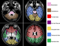 Brain regions on T1 MRI
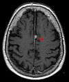 T1 (note CSF is dark) with contrast (arrow pointing to meningioma of the falx)
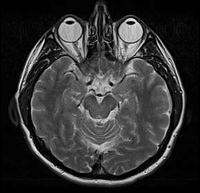
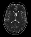 Normal axial T2-weighted MR image of the brain.
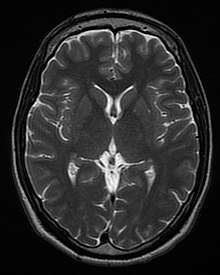
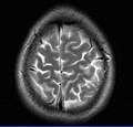 MRI image of the surface of the brain.
| Wikimedia Commons has media related to Magnetic resonance imaging of the brain. |
References
- Information, Reed Business (1978). "Britain's brains produce first NMR scans". New Scientist: 588.
- "Blood-flow checker". Popular Science: 12. 1987.
- Le Bihan D, Breton E (1987). "Method to Measure the Molecular Diffusion and/or Perfusion Parameters of Live Tissue". US Patent # 4,809,701.
- Villringer A, Rosen BR, Belliveau JW, Ackerman JL, Lauffer RB, Buxton RB, Chao YS, Wedeen VJ, Brady TJ (February 1988). "Dynamic imaging with lanthanide chelates in normal brain: contrast due to magnetic susceptibility effects". Magnetic Resonance in Medicine. 6 (2): 164–74. doi:10.1002/mrm.1910060205. PMID 3367774.
- Faro SH, Mohamed FB (2010-01-15). Bold fMRI. a guide to functional imaging for neuroscientists. Springer. ISBN 978-1-4419-1328-9. Retrieved 10 June 2015.
- Howe FA, Filler AG, Bell BA, Griffiths JR (December 1992). "Magnetic resonance neurography". Magnetic Resonance in Medicine. 28 (2): 328–38. doi:10.1002/mrm.1910280215. PMID 1461131.
- Filler AG, Howe FA, Hayes CE, Kliot M, Winn HR, Bell BA, Griffiths JR, Tsuruda JS (March 1993). "Magnetic resonance neurography". Lancet. 341 (8846): 659–61. doi:10.1016/0140-6736(93)90422-d. PMID 8095572.
- Filler A (October 2009). "Magnetic resonance neurography and diffusion tensor imaging: origins, history, and clinical impact of the first 50,000 cases with an assessment of efficacy and utility in a prospective 5000-patient study group". Neurosurgery. 65 (4 Suppl): A29–43. doi:10.1227/01.neu.0000351279.78110.00. PMC 2924821. PMID 19927075.
- Basser PJ (2010). "Invention and Development of Diffusion Tensor MRI (DT-MRI or DTI) at the NIH". Diffusion MRI. Oxford University Press. pp. 730–740. doi:10.1093/med/9780195369779.003.0047. ISBN 9780195369779.
- Hajnal JV, De Coene B, Lewis PD, Baudouin CJ, Cowan FM, Pennock JM, Young IR, Bydder GM (July 1992). "High signal regions in normal white matter shown by heavily T2-weighted CSF nulled IR sequences". Journal of Computer Assisted Tomography. 16 (4): 506–13. doi:10.1097/00004728-199207000-00002. PMID 1629405.
- Koretsky AP (August 2012). "Early development of arterial spin labeling to measure regional brain blood flow by MRI". NeuroImage. 62 (2): 602–7. doi:10.1016/j.neuroimage.2012.01.005. PMC 4199083. PMID 22245338.
- Reichenbach JR, Venkatesan R, Schillinger DJ, Kido DK, Haacke EM (July 1997). "Small vessels in the human brain: MR venography with deoxyhemoglobin as an intrinsic contrast agent". Radiology. 204 (1): 272–7. doi:10.1148/radiology.204.1.9205259. PMID 9205259.
- Mansfield P, Coxon R, Glover P (May 1994). "Echo-planar imaging of the brain at 3.0 T: first normal volunteer results". Journal of Computer Assisted Tomography. 18 (3): 339–43. doi:10.1097/00004728-199405000-00001. PMID 8188896.
- Robitaille PM, Abduljalil AM, Kangarlu A, Zhang X, Yu Y, Burgess R, Bair S, Noa P, Yang L, Zhu H, Palmer B, Jiang Z, Chakeres DM, Spigos D (October 1998). "Human magnetic resonance imaging at 8 T". NMR in Biomedicine. 11 (6): 263–5. doi:10.1002/(SICI)1099-1492(199810)11:6<263::AID-NBM549>3.0.CO;2-0. PMID 9802467.
- Vaughan T, DelaBarre L, Snyder C, Tian J, Akgun C, Shrivastava D, Liu W, Olson C, Adriany G, Strupp J, Andersen P, Gopinath A, van de Moortele PF, Garwood M, Ugurbil K (December 2006). "9.4T human MRI: preliminary results". Magnetic Resonance in Medicine. 56 (6): 1274–82. doi:10.1002/mrm.21073. PMC 4406343. PMID 17075852.
- Ebel K, Benz-Bohm G (1999). Differential diagnosis in pediatric radiology. Thieme. pp. 538–. ISBN 978-3-13-108131-5. Retrieved 18 July 2011.
- Bradley WG, Brant-Zawadzki M, Cambray-Forker J (2001-01-15). MRI of the brain. Surendra Kumar. ISBN 978-0-7817-2568-2. Retrieved 24 July 2011.
- Roos KL, Tunkel AR (2010). Bacterial infections of the central nervous system. Elsevier Health Sciences. pp. 69–. ISBN 978-0-444-52015-9. Retrieved 18 July 2011.
- American Medical Society for Sports Medicine (24 April 2014), "Five Things Physicians and Patients Should Question", Choosing Wisely: an initiative of the ABIM Foundation, American Medical Society for Sports Medicine, retrieved 29 July 2014
- Churchill Nathan W., Hutchison Michael G., Richards Doug, Leung General, Graham Simon J., Schweizer Tom A. (2017). "The first week after concussion: Blood flow, brain function and white matter microstructure". NeuroImage: Clinical. 14: 480–489. doi:10.1016/j.nicl.2017.02.015. PMC 5334547. PMID 28280686.CS1 maint: multiple names: authors list (link)
- Garel C (2004). MRI of the fetal brain: normal development and cerebral pathologies. Springer. ISBN 978-3-540-40747-8. Retrieved 24 July 2011.
- Butler P, Mitchell AW, Ellis H (2007-11-19). Applied Radiological Anatomy for Medical Students. Cambridge University Press. pp. 12–. ISBN 978-0-521-81939-8. Retrieved 18 July 2011.
- Tofts, Paul (2005-09-01). Quantitative MRI of the Brain: Measuring Changes Caused by Disease. John Wiley and Sons. pp. 86–. ISBN 978-0-470-86949-9. Retrieved 18 July 2011.
- Chowdhury R, Wilson I, Rofe C, Lloyd-Jones G (2010-04-19). Radiology at a Glance. John Wiley and Sons. pp. 95–. ISBN 978-1-4051-9220-0. Retrieved 18 July 2011.
- Granacher RP (2007-12-20). Traumatic brain injury: methods for clinical and forensic neuropsychiatric assessment. CRC Press. pp. 247–. ISBN 978-0-8493-8138-6. Retrieved 18 July 2011.
| X-ray/ Radiography |
| ||||||||||||
|---|---|---|---|---|---|---|---|---|---|---|---|---|---|
| MRI | |||||||||||||
| Ultrasound | |||||||||||||
| Radionuclide |
| ||||||||||||
| Optical/Laser | |||||||||||||
| Thermography |
| ||||||||||||
| Target conditions |
| ||||||||||||
| |||||||||||||