Lobes of the brain
The lobes of the brain were originally a purely anatomical classification, but have been shown also to be related to different brain functions. The cerebrum, the largest portion of the human brain, is divided into lobes, but so is the cerebellum. If not specified, the expression "lobes of the brain" refers to the cerebrum.
| Cerebral lobes | |
|---|---|
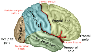 Lateral surface of cerebrum. 4 lobes are shown. | |
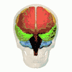
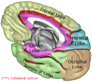 Medial surface of cerebrum. 5 lobes are shown. | |
| Identifiers | |
| NeuroNames | 1210 |
| NeuroLex ID | birnlex_922 |
| TA | A14.1.09.005 |
| FMA | 77800 |
| Anatomical terms of neuroanatomy | |
Terminologia Anatomica (1998) and Terminologia Neuroanatomica (2017) divides the cerebrum into 6 lobes.[1][2]
Frontal lobe
The frontal lobe is located at the front of each cerebral hemisphere and positioned in front of the parietal lobe and above and in front of the temporal lobe. It is separated from the parietal lobe by a space between tissues called the central sulcus, and from the temporal lobe by a deep fold called the lateral sulcus also called the Sylvian fissure. The precentral gyrus, forming the posterior border of the frontal lobe, contains the primary motor cortex, which controls voluntary movements of specific body parts.
The frontal lobe contains most of the dopamine-delicate neurons in the cerebral cortex. The dopamine system is associated with reward, attention, short-term memory tasks, planning, and motivation. Dopamine tends to limit and select sensory information arriving from the thalamus to the forebrain. A report from the National Institute of Mental Health says a gene variant that reduces dopamine activity in the prefrontal cortex is related to poorer performance and inefficient functioning of that brain region during working memory tasks, and to a slightly increased risk for schizophrenia.[3]
Parietal lobe
The parietal lobe is positioned above the occipital lobe and behind the frontal lobe and central sulcus.
The parietal lobe integrates sensory information among various modalities, including spatial sense and navigation (proprioception), the main sensory receptive area for the sense of touch (mechanoreception) in the somatosensory cortex which is just posterior to the central sulcus in the postcentral gyrus,[4] and the dorsal stream of the visual system. The major sensory inputs from the skin (touch, temperature, and pain receptors), relay through the thalamus to the parietal lobe.
Several areas of the parietal lobe are important in language processing. The somatosensory cortex can be illustrated as a distorted figure — the homunculus (Latin: "little man"), in which the body parts are rendered according to how much of the somatosensory cortex is devoted to them.[5] The superior parietal lobule and inferior parietal lobule are the primary areas of body or spatial awareness. A lesion commonly in the right superior or inferior parietal lobule leads to hemineglect.
Occipital lobe
The occipital lobe is the visual processing center of the mammalian brain containing most of the anatomical region of the visual cortex.[6] The primary visual cortex is Brodmann area 17, commonly called V1 (visual one). Human V1 is located on the medial side of the occipital lobe within the calcarine sulcus; the full extent of V1 often continues onto the posterior pole of the occipital lobe. V1 is often also called striate cortex because it can be identified by a large stripe of myelin, the Stria of Gennari. Visually driven regions outside V1 are called extrastriate cortex. There are many extrastriate regions, and these are specialized for different visual tasks, such as visuospatial processing, color differentiation, and motion perception.
Temporal lobe
The temporal lobe is located beneath the lateral fissure on both cerebral hemispheres of the mammalian brain.[7]
The temporal lobe is involved in processing sensory input into derived meanings for the appropriate retention of visual memories, language comprehension, and emotion association.[8]:21
Limbic lobe
The limbic lobe is an arc-shaped region of cortex on the medial surface of each cerebral hemisphere of the mammalian brain, consisting of parts of the frontal, parietal and temporal lobes. The term is ambiguous, with some authors including the paraterminal gyrus, the subcallosal area, the cingulate gyrus, the parahippocampal gyrus, the dentate gyrus, the hippocampus and the subiculum;[9] while the Terminologia Anatomica includes the cingulate sulcus, the cingulate gyrus, the isthmus of cingulate gyrus, the fasciolar gyrus, the parahippocampal gyrus, the parahippocampal sulcus, the dentate gyrus, the fimbrodentate sulcus, the fimbria of hippocampus, the collateral sulcus, and the rhinal sulcus, and omits the hippocampus.
Insular cortex
The insular cortex is a portion of the cerebral cortex folded deep within the lateral sulcus (the fissure separating the temporal lobe from the parietal and frontal lobes).
The insulae are believed to be involved in consciousness and play a role in diverse functions usually linked to emotion or the regulation of the body's homeostasis. These functions include perception, motor control, self-awareness, cognitive functioning, and interpersonal experience. In relation to these, it is involved in psychopathology.
The insular cortex is divided into two parts: the larger anterior insula and the smaller posterior insula in which more than a dozen field areas have been identified. The cortical area overlying the insula toward the lateral surface of the brain is the operculum (meaning lid). The opercula are formed from parts of the enclosing frontal, temporal, and parietal lobes.
Additional images
 Interior view of brain.
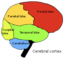
 Brain lobes. Colorings are same as the left and
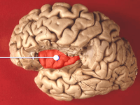
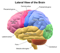 Lateral View of the Brain.
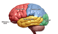 Brain Lobes.
References
- Guilherme Carvalhal Ribas (2010). “The Cerebral Sulci and Gyri”. Neurosurg Focus 56 (2): E2. PMID 20121437.
- FIPAT. Terminologia Neuroanatomica. FIPAT.library.dal.ca. Federative International Programme for Anatomical Terminology, February 2017
- "Gene Slows Frontal Lobes, Boosts Schizophrenia Risk". National Institute of Mental Health. May 29, 2001. Archived from the original on April 4, 2015. Retrieved 2013-06-20.
- http://www.ruf.rice.edu/~lngbrain/cglidden/parietal.html
- Schacter, D. L., Gilbert, D. L. & Wegner, D. M. (2009). Psychology. (2nd ed.). New Work (NY): Worth Publishers.
- "SparkNotes: Brain Anatomy: Parietal and Occipital Lobes". Archived from the original on 2007-12-31. Retrieved 2008-02-27.
- "Temporal Lobe". Langbrain. Rice University. Retrieved 2 January 2011.
- Smith; Kosslyn (2007). Cognitive Psychology: Mind and Brain. New Jersey: Prentice Hall. pp. 21, 194–199, 349.
- Fix, JD (2008). "Gross anatomy of the brain". Neuroanatomy (fourth ed.). Philadelphia: Lippincott Williams & Wilkins. p. 6. ISBN 0-7817-7245-1.