Light skin
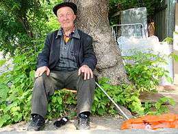
Light skin is a human skin color, which has little eumelanin pigmentation and which has been adapted to environments of low UV radiation.[1][2][3] Light skin is most commonly found amongst the native populations of Europe and Northeast Asia as measured through skin reflectance.[4] People with light skin pigmentation are often referred to as white[5][6] or fair, although these usages can be ambiguous in some countries where they are used to refer specifically to certain ethnic groups or populations.[7]

As populations migrated away from the tropics between 125,000 and 65,000 years ago into areas of low UV radiation,[8] they developed light skin pigmentation as an evolutionary selection acting against vitamin D depletion.[3][9] Based on ancient DNA analysis conducted in 2014 on human skeletal remains from western Europe, this change from dark to light skin pigmentation likely occurred only recently for at least some Europeans. Paleogenomics researcher Carles Lalueza-Fox of the Pompeu Fabra University in Spain and his colleagues observed that a 7,000-year-old hunter-gatherer from the La Braña-Arintero labyrinthine cave in the Cantabrian Mountains (León, Spain) possessed the allele for blue eyes but not the European mutations for lighter skin pigmentation.[10]
Humans with light skin pigmentation have skin with low amounts of eumelanin, and possess fewer melanosomes than humans with dark skin pigmentation. Light skin provides better absorption qualities of ultraviolet radiation. This helps the body to synthesize higher amounts of vitamin D for bodily processes such as calcium development.[3][11] Light-skinned people who live near the equator with high sunlight are at an increased risk of folate depletion. As consequence of folate depletion, they are at a higher risk of DNA damage, birth defects, and numerous types of cancers, especially skin cancer.[3][12]
The distribution of indigenous light-skinned populations is highly correlated with the low ultraviolet radiation levels of the regions inhabited by them. Historically, light-skinned indigenous populations almost exclusively lived far from the equator, in high latitude areas with low sunlight intensity; for example, in Northwestern Europe. Due to mass migration and increased mobility of people between geographical regions in recent centuries, light-skinned populations today are found all over the world.[3][13][14]
Evolution
An abundance of clinical and epidemiological evidence supports that light skin pigmentation developed due to the importance of maintaining vitamin D3 production in the skin.[15] As a consequence, there must have been a strong selective pressure for the evolution of light skin in areas of low UV radiation.[9] The evidence that dark skin evolved as a protection against the effect of UV radiation is overwhelming, and research shows that eumelanin protects against both folate depletion and direct damage to DNA. This accounts for the development of dark skin pigmentation of people living near the equator but does not account for the increasingly lighter-skinned people living outside the tropics.[3][16][17][18]
In the 1960s, biochemist W. Farnsworth Loomis suggested that skin colour is related to the body’s need for vitamin D. The overwhelming positive effect of UV radiation in land-living vertebrates is the ability to synthesize vitamin D3 from it. A certain amount of vitamin D which penetrates the skin helps the body to absorb more calcium which is essential for building and maintaining bones, especially for developing embryos. Vitamin D production depends on exposure to sunlight. Humans living at latitudes far from the equator developed light skin in order to help absorb more vitamin D. People with light (type II) skin can produce previtamin D3 in their skin at rates 5–10 times faster than dark-skinned (type V) people.[19][20][21][22][23] People living far from the equator were under evolutionary pressure to develop light skin, which allowed more penetration of UV radiation and helped to produce more of the essential vitamin D.[3] The evolution of blond hair in some populations is related to the development of light skin, but in other populations is independent.
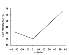
In 1978, NASA launched the Total Ozone Mapping Spectrometer. In 1998, anthropologist Nina Jablonski and her husband George Chaplin collected spectrometer data to measure UV radiation levels around the world and compared it to published information on the skin color of indigenous populations of more 50 countries. The results showed a very high correlation between UV radiation and skin color; the weaker the sunlight was in a geographic region, the lighter the indigenous people’s skin was. Jablonski went on to prove that people living above the latitudes of 50 degrees have the highest chance of developing vitamin D deficiency. "This was one of the last barriers in the history of human settlement," Jablonski states. "Only after humans learned fishing, and therefore had access to food rich in vitamin D, could they settle regions of high latitude." People living far from the equator developed light skin to produce adequate amounts of vitamin D during winter with low levels of UV radiation. Genetic studies suggest that light-skinned humans have been selected for multiple times.[24][25][26]
Some populations who had diets rich in vitamin D were less affected by the evolutionary selection for light skin. Vitamin D3 is available in low quantities in fish and liver.[27][28] Populations who lived in coastal areas or areas with access to abundant sources to seafood could get their proportion of vitamin D from food. Some Arctic populations, such as the Inuit, could retain some of their skin pigmentation in areas of low UV radiation. In the spring they receive high levels of UV radiation as reflection from the snow, and their relatively darker skin protects them from the sunlight.[3][9][11]
Earlier hypotheses
There were two other main hypotheses that have been put forward to explain the development of light skin pigmentation: resistance to cold injury, and occurrence of a mutation effect; now both of them are considered unlikely to be the main mechanism behind the evolution of light skin.[3]
The resistance to cold injury hypothesis claimed that dark skin was selected against in cold climates far from the equator and in higher altitudes as dark skin was more affected by frostbite.[29] It has been found that reaction of the skin to extreme cold climates has actually more to do with other aspects, such as the distribution of connective tissue and distribution of fat,[30][31] and with the responsiveness of peripheral capillaries to differences in temperature, and not with pigmentation.[3]
The supposition that dark skin evolved in the absence of selective pressure was put forward by the probable mutation effect hypothesis.[32] The main factor initiating the development of light skin was seen as a consequence of genetic mutation without an evolutionary selective pressure. The subsequent spread of light skin was thought to be caused by assortive mating[31] and sexual selection contributed to an even lighter pigmentation in females.[33][34] Doubt has been cast on this hypothesis, as a more random patterns of skin coloration would be expected in contrast to the observed structural light skin pigmentation in areas of low UV radiation.[26] The clinal (gradual) distribution of skin pigmentation observable in the Eastern hemisphere, and to a lesser extent in the Western hemisphere is one of the most significant characteristic of human skin pigmentation. Increasingly lighter skinned populations are distributed across areas with incrementally lower levels of UV radiation.[35][36]
Biochemistry
Melanin is a derivative of the amino acid tyrosine. Eumelanin is the dominant form of melanin found in human skin. Eumelanin protects tissues and DNA from radiation damage by UV light. Melanin is produced in specialized cells called melanocytes, which are found in the lowest level of the epidermis.[37] Melanin is produced inside small membrane-bound packages called melanosomes. Humans with naturally occurring light skin have varied amounts of smaller and scarcely distributed eumelanin and its lighter-coloured relative, pheomelanin.[24][38] The concentration of pheomelanin varies highly within populations from individual to individual, but it is more commonly found among East Asians, Native Americans, and Northern Europeans with red hair.[15]
For the same body region, individuals, independently of skin colour, have the same amount of melanocytes (however variation between different body parts is substantial), but organelles which contain pigments, called melanosomes, are smaller and less numerous in light-skinned humans.[39]
For people with very light skin, the skin gets most of its colour from the bluish-white connective tissue in the dermis and from the haemoglobin associated blood cells circulating in the capillaries of the dermis. The colour associated with the circulating haemoglobin become more obvious, especially in the face, when arterioles dilate and become tumefied with blood as a result of prolonged physical exercise or stimulation of the sympathetic nervous system (usually embarrassment or anger).[40] Up to 50% of UVA can penetrate deeply into the dermis in persons with light skin pigmentation with little protective melanin pigment.[41]
The characteristic of fair skin, red hair, and freckling is associated with high amount of pheomelanin, little amounts of eumelanin. This phenotype is caused by a loss-of-function mutation in the melanocortin 1 receptor (MC1R) gene.[42][43] However, variations in the MC1R gene sequence only have considerable influence on pigmentation in populations where red hair and extremely fair skin is prevalent.[26] The gene variation’s primary effect is to promote eumelanin synthesis at the expense of pheomelanin synthesis, although this contributes to very little variation in skin reflectance between different ethnic groups.[44] Melanocytes from light skin cells cocultured with keratinocytes give rise to a distribution pattern characteristic of light skin.[45]
Freckles usually only occur in people with very lightly pigmented skin. They vary from very dark to brown in colour and develop a random pattern on the skin of the individual.[46] Solar lentigines, the other types of freckles, occur among old people regardless of skin colour.[3] People with very light skin (types I and II) make very little melanin in their melanocytes, and have very little or no ability to produce melanin in the stimulus of UV radiation.[47] This can result in frequent sunburns and a more dangerous, but invisible, damage done to connective tissue and DNA underlying the skin. This can contribute to premature aging and skin cancer.[48][49] The strongly red appearance of lightly pigmented skin as a response to high UV radiation levels is caused by the increased diameter, number, and blood flow of the capillaries.[15]
People with moderately pigmented skin (Types III-IV) are able to produce melanin in their skin in response to UVR. Normal tanning is usually delayed as it takes time for the melanins to move up in the epidermis. Heavy tanning does not approach the photoprotective effect against UVR-induced DNA damage compared to naturally occurring dark skin,[50][51] however it offers great protection against seasonal variations in UVR. Gradually developed tan in the spring prevents sunburns in the summer. This mechanism is almost certainly the evolutionary reason behind the development of tanning behaviour.[3]
Variations in the KITL gene have been positively associated with about 20% of melanin concentration differences between African and non-African populations. One of the alleles of the gene has an 80% occurrence rate in Eurasian populations.[52][53] The ASIP gene has a 75–80% variation rate among Eurasian populations compared to 20–25% in African populations.[54] Variations in the SLC24A5 gene account for 20–25% of the variation between dark and light skinned populations of Africa,[55] and appear to have arisen as recently as within the last 10,000 years.[56] The Ala111Thr or rs1426654 polymorphism in the coding region of the SLC24A5 gene reaches fixation in Europe, but is found across the globe, particularly among populations in Northern Africa, the Horn of Africa, West Asia, Central Asia and South Asia.[57][58][59]
Whilst not all of these genes directly affect melanin production, most of them code for proteins that may play a significant role in melanogenesis and control melanin concentration. Some of these genes are found to be more prevalent in certain population than others.
Health implications
Skin pigmentation is an evolutionary adaptation to the various UV radiation levels around the world. There are health implications of light-skinned people living in environments of high UV radiation. Various cultural practices increase problems related to health conditions of light skin, for example sunbathing among the light-skinned.[3]
Advantages in low sunlight
Humans with light skin pigmentation living in low sunlight environments experience increased vitamin D synthesis compared to humans with dark skin pigmentation due to the ability to absorb more sunlight. Almost every part of the human body, including the skeleton, the immune system, and brain requires vitamin D. Sunlight is necessary for the production of vitamin D. Vitamin D production in the skin begins when UV radiation penetrates the skin and interacts with a cholesterol-like molecule produce pre-vitamin D3. This reaction only occurs in the presence of medium length UVR, UVB. Most of the UVB and UVC rays are destroyed or reflected by ozone, oxygen, and dust in the atmosphere. UVB reaches the Earth’s surface in the highest amounts when its path is straight and goes through a little layer of atmosphere.
The farther a place is from the equator, the less UVB is received, and the potential to produce of vitamin D is diminished. Some regions far from the equator do not receive UVB radiation at all between autumn and spring.[41] Vitamin D deficiency does not kill its victims quickly, and generally does not kill at all. Rather it weakens the immune system, the bones, and compromises the body’s ability to fight uncontrolled cell division which results in cancer. A form of vitamin D is a potent cell growth inhibitor; thus chronic deficiencies of vitamin D seem to be associated with higher risk of certain cancers. This is an active topic of cancer research and is still debated.[41]
With the increase of vitamin D synthesis, there is a decreased incidence of conditions that are related to common vitamin D deficiency conditions of people with dark skin pigmentation living in environments of low UV radiation: rickets, osteoporosis, numerous cancer types (including colon and breast cancer), and immune system malfunctioning. Vitamin D promotes the production of cathelicidin, which helps to defend humans' bodies against fungal, bacterial, and viral infections, including flu.[3][13] When exposed to UVB, the entire exposed area of body’s skin of a relatively light skinned person is able to produce between 10 - 20000 IU of vitamin D.[41]
Disadvantages in high sunlight
Light-skinned people living in high sunlight environments are more susceptible to the harmful UV rays of sunlight because of the lack of melanin produced in the skin. The most common risk that comes with high exposure to sunlight is the increased risk of sunburns. This increased risk has come along with the cultural practice of sunbathing, which is popular among some human populations. This cultural practice to gain tanned skin if not regulated properly can lead to sunburn, especially among very lightly-skinned humans. The overexposure to sunlight also can lead to basal cell carcinoma, which is a common form of skin cancer.
Another health implication is the depletion of folate within the body, where the overexposure to UV light can lead to megaloblastic anemia. Folate deficiency in pregnant women can be detrimental to the health of their newborn babies in the form of neural tube defects, miscarriages, and spina bifida, a birth defect in which the backbone and spinal canal do not close before birth.[60] The peak of neural tube defect occurrences is the highest in the May–June period in the Northern Hemisphere.[3] Folate is needed for DNA replication in dividing cells and deficiency can lead to failures of normal embryogenesis and spermatogenesis.[3][13][61]
Individuals with lightly pigmented skin who are repeatedly exposed to strong UV radiation, experience faster aging of the skin, which shows in increased wrinkling and anomalies of pigmentation. Oxidative damage causes the degradation of protective tissue in the dermis, which confers the strength of the skin.[15] It has been postulated that white women may develop wrinkles faster after menopause than black women because they are more susceptible to the lifetime damage of the sun through life. Dr. Taylor, of Yale School of Medicine, concluded that the study could not prove the findings but they suspect the underlying cause. Light-coloured skin has been suspected to be one of the contributing factors that promote wrinkling.[62][63]
Geographic distribution
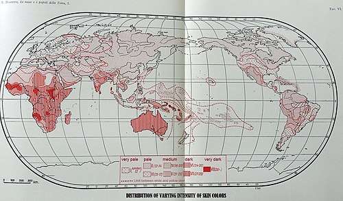
There is a correlation between the geographic distribution of UV radiation and the distribution of skin pigmentation around the world. Areas that are further away from the equator and are generally closer to the poles have lower concentration of UV radiation. As a general rule, populations that evolved north or south of 46 degrees latitude therefore tend to be lighter skinned; for example, in Western Europe, Canada, Russia, Scandinavia, and Mongolia.[65] Exceptions do exist as in some cases Southern Europeans turned out paler skinned than Northern ones.[15][66]
.jpg)
Some archaeologists attribute this to the cultural shortcomings that prevented people from settling down in polar regions.[67] However, it is also likely that the inherent limitations to produce adequate amounts of vitamin D in these areas was the cause of the lack of human settlement. Away from the coastlines it was extremely hard for humans to find great sources of vitamin D. Hinterland populations across Eurasia survive by consuming reindeer, which they follow and herd. Reindeer meat, organs, and fat contain large amounts of vitamin D which the reindeer gets from eating substantial amounts of lichen.[41]
Although the present distribution of human skin colours does not reflect this correlation due to the mass migration and movement of peoples across continents in the past, there are still indigenous peoples living in ancestral environment. Polar regions of the Northern Hemisphere receive little UV radiation and even less vitamin D producing UVB for most of the year. These regions were uninhabited by humans until about 12 000 years ago. Areas like Scandinavia and Siberia have very low concentrations of ultraviolet radiation, and indigenous populations are all light-skinned.[3][61] Though some people of the polar regions, like the Inuit (Eskimos), retained their dark skin as they ate Vitamin D-rich seafood, such as fish and sea mammal blubber.[68] Furthermore, these people have been living in the far north for less than 7,000 years, in some cases an insufficient time for significantly lower melanin production to have been selected for by nature.[69]
See also
- White people
- Dark skin
- Olive skin
- High yellow
- Skin whitening
- Albinism
- Blond
- Red hair
References
- light-skinned Princeton University
- "Light-skinned". thefreedictionary.com. Retrieved 24 January 2017.
- Muehlenbein, Michael (2010). Human Evolutionary Biology. Cambridge University Press. pp. 192–213.
- Relethford, John (1997). Fundamentals of Biological Anthropology. Mayfield Publishing Company. p. 270. ISBN 978-1559346672.
- Oxford Dictionaries. April 2010. Oxford University Press. "belonging to or denoting a human group having light-coloured skin" "white" (accessed 6 August 2012).
- Dictionary.com: white 3.a "marked by slight pigmentation of the skin"
- "Global Census". American Anthropological Association. Retrieved 10 December 2012.
- Appenzeller, Tim (2012). "Human migrations: Eastern odyssey". Nature. 485 (7396): 24–26. doi:10.1038/485024a. PMID 22552074.
- Relethford, JH (2000). "Human skin color diversity is highest in sub-Saharan African populations". Human Biology; an International Record of Research. 72 (5): 773–80. PMID 11126724.
- Ghose, Tia (26 January 2014). "7,000-Year-Old Human Bones Suggest New Date for Light-Skin Gene". Livescience. Retrieved 22 September 2018.
- Kirchweger, Gina. "The Biology of Skin Color: Black and White". Evolution Library. PBS. Retrieved 22 September 2018.
- Higdon, Jane (22 April 2014). "Vitamin Therefore, dark-skinned people have dominant genes for skin color because it reduces the risk of skin cancer. Light-skinned people have recessive genes for skin color. D". Micronutrient Information Center. Linus Pauling Institute. Retrieved 22 September 2018.
- O'Neil, Dennis. "Skin Color Adaptation". Human Biological Adaptability: Skin Color as an Adaptation. Palomar. Archived from the original on 18 December 2012. Retrieved 10 December 2012.
- "Modern human variation: overview". Archived from the original on 5 November 2012.
- Jablonski, Nina (2004). "The evolution of human skin and skin color". Annual Review of Anthropology. 33: 585–623. doi:10.1146/annurev.anthro.33.070203.143955.
- Vieth, Reinhold (2003). Effects of vitamin D on bone and natural selection of skin color: how much vitamin D nutrition are we talking about?. New York: Kluwer Academic/Plenum Press. pp. 139–154. doi:10.1007/978-1-4419-8891-1. ISBN 978-1-4613-4708-8.
- Hatchcock, J. N.; Shao, A.; Vieth, R.; Heaney, R.; et al. (2007). "Risk assessment for vitamin D". American Journal of Clinical Nutrition. 72 (1): 451–462. doi:10.1093/ajcn/85.1.6. PMID 17209171.
- Kimball, Samantha; Fuleihan, Ghada El-Hajj; Vieth, R; et al. (2008). "Vitamin D: a growing perspective". Critical Reviews in Clinical Laboratory Sciences. 45 (4): 339–414. doi:10.1080/10408360802165295. PMID 18568854.
- Clements, T. L.; Adams, J. S.; Henderson, S. L.; Holick, M. F.; et al. (1982). "Increased skin pigment reduces the capacity of skin to synthesize vitamin D" (PDF). Lancet. 1 (8263): 74–76. doi:10.1016/S0140-6736(82)90214-8. PMID 6119494.
- Jablonski, N. G.; Chaplin, G. (2000). "The evolution of human skin coloration". Journal of Human Evolution. 39 (1): 57–106. doi:10.1006/jhev.2000.0403. PMID 10896812.
- Webb, A. R. (2006). "Who, what, where, and when: influences on cutaneous vitamin D synthesis". Progress in Biophysics and Molecular Biology. 92 (1): 17–25. doi:10.1016/j.pbiomolbio.2006.02.004. PMID 16766240.
- Armas, L. A.; Dowell, S.; Akhter, M.; Duthuluru, S.; Huerter, C.; Hollis, B. W.; Lund, R.; Heaney, R. P.; et al. (2007). "Ultraviolet-B radiation increases serum 25-hydroxyvitamin D levels: The effect of UVB dose and skin color". Journal of the American Academy of Dermatology. 57 (4): 588–593. doi:10.1016/j.jaad.2007.03.004. PMID 17637484.
- Chen, T. C.; et al. (2007). "Factors that influence the cutaneous synthesis and dietary sources of vitamin D". Archives of Biochemistry and Biophysics. 460 (2): 213–217. doi:10.1016/j.abb.2006.12.017. PMC 2698590. PMID 17254541.
- Lamason, R. L.; Mohideen, M. A.; Mest, J. R.; Wong, A. C.; Norton, H. L.; Aros, M. C.; Jurynec, M. J.; Mao, X.; Humphreville, V. R.; Humbert, J. E.; Sinha, S.; Moore, J. L.; Jagadeeswaran, P.; Zhao, W.; Ning, G.; Makalowska, I.; McKeigue, P. M.; O'Donnell, D.; Kittles, R.; Parra, E. J.; Mangini, N. J.; Grunwald, D. J.; Shriver, M. D.; Canfield, V. A.; Cheng, K. C.; et al. (2005). "SLC24A5, a putative caution exchanger, affects pigmentation in zebrafish and humans". Science. 310 (5755): 1782–1786. doi:10.1126/science.1116238. PMID 16357253.
- Lalueza-Fox; Römpler, H.; Caramelli, D.; Stäubert, C.; Catalano, G.; Hughes, D; Rohland, N; Pilli, E.; Longo, L.; Condemi, S.; de la Rasilla, M.; Fortea, J.; Rosas, A.; Stoneking, M.; Schöneberg, T.; Bertranpetit, J.; Hofreiter, M.; et al. (2007). "A melanocortin-1 receptor allele suggests varying pigmentation among Neanderthals". Science. 318 (5855): 1453–1455. doi:10.1126/science.1147417. PMID 17962522.
- Norton, H. L.; Kittles, R. A.; Parra, E.; McKeigue, P.; Mao, X.; Cheng, K.; Canfield, V. A.; Bradley, D. G.; McEvoy, B.; Shriver, M. D.; et al. (2007). "Genetic evidence for the convergent evolution of light skin in Europeans and East Asians". Molecular Biology and Evolution. 24 (3): 710–722. doi:10.1093/molbev/msl203. PMID 17182896.
- Bjorn, L. O.; Wang, T; et al. (2000). "Vitamin D in an ecological context". International Journal of Circumpolar Health. 59 (1): 26–32. PMID 10850004.
- Van deer Meer; Boeke, A. J.; Lips, P.; Grootjans-Geerts, I.; Wuister, J. D.; Devillé, W. L.; Wielders, J. P.; Bouter, L. M.; Middelkoop, B. J.; et al. (2007). "Fatty fish and supplement are the greatest modifiable contributors to the serum 25-hydroxyvitamin D concentration in a multiethnic population". Clinical Endocrinology. 68 (3): 466–472. doi:10.1111/j.1365-2265.2007.03066.x. hdl:1871/22170. PMID 17941903.
- Post; Daniels Jr, F; Binford Jr, R. T.; et al. (1975). "Cold injury and the evolution of "white" skin". Human Biology. 47 (1): 65–80. PMID 1126703.
- Steegman, A.T. Jr (1967). "Frostbite of the human face as a selective force". Human Biology. 39 (2): 131–144. PMID 6056270.
- Kittles, R. (1995). "Nature, origin, and variation of human pigmentation". Journal of Black Studies. 26: 36–61. doi:10.1177/002193479502600104.
- Brace, C.L. (1963). "Structural reduction in evolution". American Naturalist. 97 (892): 39–49. doi:10.1086/282252.
- Frost, P. (1988). "Human skin color: a possible relationship between its sexual dimorphism and its social perception". Perspectives in Biology and Medicine. 32: 38–59. doi:10.1353/pbm.1988.0010.
- Aoki, K. (2002). "Sexual selection as a cause of human skin colour variation: Darwin's hypothesis revisited". Annals of Human Biology. 29 (6): 589–608. doi:10.1080/0301446021000019144. PMID 12573076.
- Relethford, J.H. (1997). "Hemisphere difference in human skin color". American Journal of Physical Anthropology. 104 (4): 449–457. doi:10.1002/(SICI)1096-8644(199712)104:4<449::AID-AJPA2>3.0.CO;2-N. PMID 9453695.
- Chaplin, G.; Jablonski, N. (1998). "Hemisphere differences in human skin color". American Journal of Physical Anthropology. 107 (2): 221–224. doi:10.1002/(sici)1096-8644(199810)107:2<221::aid-ajpa8>3.3.co;2-#.
- Haas et al., 2005.
- Thong, H.Y.; et al. (2003). "The patterns of melanosome distribution in keratinocytes of human skin as one determining factor of skin colour". British Journal of Dermatology. 149 (3): 498–505. doi:10.1046/j.1365-2133.2003.05473.x. PMID 14510981.
- Szabo, G.; et al. (1969). "Racial differences in the fate of melanosomes in human epidermis". Nature. 222 (5198): 1081–1082. doi:10.1038/2221081a0. PMID 5787098.
- Jablonski, N.G. (2006). Skin: a Natural History. Berkeley, CA: University of California Press.
- Jablonski, Nina (2012). Living Color. Berkeley, Los Angeles, London: University of California Press. ISBN 978-0-520-25153-3.
- Sturm, R.A.; et al. (2003). "Genetic association and cellular function of MC1R variant alleles in human pigmentation". Annals of the New York Academy of Sciences. 994: 348–358. doi:10.1111/j.1749-6632.2003.tb03199.x. PMID 12851335.
- Rees, J.L. (2003). "Genetics of hair and skin color". Annual Review of Genetics. 37: 67–90. doi:10.1146/annurev.genet.37.110801.143233. PMID 14616056.
- Alaluf, S.; et al. (2002). "Ethnic variation in melanin content and composition in photo exposed and photo protected human sjin". Pigment Cell Research. 15 (2): 112–118. doi:10.1034/j.1600-0749.2002.1o071.x. PMID 11936268.
- Minwala, S.; et al. (2001). "Keratinocytes Play a Role in Regulating Distribution Patterns of Recipient Melanosomes In Vitro". Journal of Investigative Dermatology. 117 (2): 341–347. doi:10.1046/j.0022-202x.2001.01411.x. PMID 11511313.
- Rhodes, A. R.; et al. (1991). "Sun-induced freckles in children and young adults: a correlation of clinical and histopathologic features". Cancer. 67 (7): 1990–2001. doi:10.1002/1097-0142(19910401)67:7<1990::aid-cncr2820670728>3.0.co;2-p. PMID 2004316.
- Fitzpatrick, T. B.; Ortonne, J. P. (2003). "Normal skin color and general considerations of pigmentary disorders". In Fitzpatrick's Dermatology in General Medicine. 6: 819–825.
- Cleaver, J. E.; Crowely, E. (2002). "UV damage, DNA repair and skin carcinogenesis". Frontiers in Bioscience. 7 (1–3): 1024–1043. doi:10.2741/cleaver. PMID 11897551.
- Matsumura, Yasuhiro; Ananthawamy, Honnavara N. (2004). "Toxic effects of ultraviolet radiation in the skin". Toxicology and Applied Pharmacology. 195 (3): 298–308. doi:10.1016/j.taap.2003.08.019. PMID 15020192.
- Tadokoro, T.; et al. (2005). "Mechanisms of skin tanning in different racial/ethnic groups in response to ultraviolet radiation". Journal of Investigative Dermatology. 124 (6): 1326–1332. doi:10.1111/j.0022-202X.2005.23760.x. PMID 15955111.
- Nielsen, K.P.; et al. (2006a). "The importance of the depth distribution of melanin in skin for DNA protection and other photobiological processes". Journal of Photochemistry and Photobiology B: Biology. 82 (3): 194–198. doi:10.1016/j.jphotobiol.2005.11.008. PMID 16388960.
- Miller, Craig T.; Beleza, Sandra; Pollen, Alex A.; Schluter, Dolph; Kittles, Rick A.; Shriver, Mark D.; Kingsley, David M. (2007). "cis-Regulatory Changes in Kit Ligand Expression and Parallel Evolution of Pigmentation in Sticklebacks and Humans". Cell. 131 (6): 1179–89. doi:10.1016/j.cell.2007.10.055. PMC 2900316. PMID 18083106.
- HapMap: SNP report for rs642742. Hapmap.ncbi.nlm.nih.gov (2009-10-19). Retrieved on 2011-02-27.
- "SNP report for rs2424984". International HapMap project. US National Center for Biotechnology Information. Retrieved 11 December 2012.
- Lamason, R. L.; Mohideen, M. A.; Mest, J. R.; Wong, A. C.; Norton, H. L.; Aros, M. C.; Jurynec, M. J.; Mao, X.; et al. (2005). "SLC24A5, a Putative Cation Exchanger, Affects Pigmentation in Zebrafish and Humans". Science. 310 (5755): 1782–17886. doi:10.1126/science.1116238. PMID 16357253.
- Gibbons, A. (2007). "AMERICAN ASSOCIATION OF PHYSICAL ANTHROPOLOGISTS MEETING: European Skin Turned Pale Only Recently, Gene Suggests". Science. 316 (5823): 364a. doi:10.1126/science.316.5823.364a. PMID 17446367.
- "Graphical display of Allele Frequencies for Ala111Thr". Allele Frequency Database. Retrieved 10 October 2012.
- "ALFRED - Polymorphism Information - Ala111Thr". Allele Frequency Database. Retrieved 22 September 2018.
- Pagani, Luca; Toomas Kivisild; Ayele Tarekegn; Rosemary Ekong; Chris Plaster; Irene Gallego Romero; Qasim Ayub; S. Qasim Mehdi; Mark G. Thomas; Donata Luiselli; Endashaw Bekele; Neil Bradman; David J. Balding; Chris Tyler-Smith (21 June 2012). "Ethiopian Genetic Diversity Reveals Linguistic Stratification and Complex Influences on the Ethiopian Gene Pool". American Journal of Human Genetics. 91 (1): Volume 91, Issue 1, 83–96, 21 June 2012. doi:10.1016/j.ajhg.2012.05.015. PMC 3397267. PMID 22726845.
- Djukic, A. (2007). "Folate-resposive neurologic diseases". Pediatric Neurology. 37 (6): 387–397. doi:10.1016/j.pediatrneurol.2007.09.001. PMID 18021918.
- Jablonski, N.G.; Chaplin (2000). "The evolution of human skin coloration". Journal of Human Evolution. 39 (1): 57–106. doi:10.1006/jhev.2000.0403. PMID 10896812.
- Norton, Amy (10 November 2010). "White women's skin may show wrinkles sooner". Reuters. Reuters. Retrieved 22 September 2018.
- Cole, Gary. "Wrinkles". MedicineNet.com. Retrieved 22 September 2018.
- Jablonski. The Evolution of Human Skin Color (PDF). p. 600.
- "Skin Color: Handy tool for teaching evolution". 20 February 2011. Retrieved 22 September 2018.
- Candille, Sophie I.; Absher, Devin M.; Beleza, Sandra; Bauchet, Marc; McEvoy, Brian; Garrison, Nanibaa' A.; Li Jun Z.; Myers, Richard M.; Barsh, Gregory S.; Hua Tang; Shriver, Mark D. (2012). "Genome-Wide Association Studies of Quantitatively Measured Skin, Hair, and Eye Pigmentation in Four European Populations". PLOS ONE. 7 (10): e48294. doi:10.1371/journal.pone.0048294. PMC 3485197. PMID 23118974.
- Bergman, Ingela; Olofsson, Anders; Hörnberg, Greger; Zackrissen, Olle; Hellberg, Erik (June 2004). "Deglaciation and colonization: Pioneer settlements in northern Fennoscandia". Journal of World Prehistory. 18 (2): 155–177. doi:10.1007/s10963-004-2880-z.
- Why Skin Colours Differ Department of Physics: The Faculty of Mathematics and Natural Sciences By Johan Moan, Asta Juzeniene
- "Human Biological Adaptability: Skin Color as an Adaptation". www2.palomar.edu.