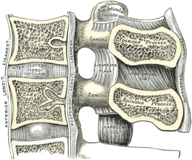
Ligamenta flava
The ligamenta flava (singular, ligamentum flavum, Latin for yellow ligament) are a series of ligaments that connect the ventral parts of the laminae of adjacent vertebrae. Each ligamentum flavum connects two adjacent vertrebrae, beginning with the junction of the axis and third cervical vertebra, continuing down to the junction of the fifth lumbar vertebra and the sacrum.[1] They are best seen from the interior of the vertebral canal; when looked at from the outer surface they appear short, being overlapped by the lamina of the vertebral arch.
| Ligamenta flava | |
|---|---|
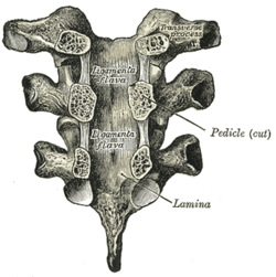 Vertebral arches of three thoracic vertebrae viewed from the front | |
 | |
| Details | |
| Identifiers | |
| Latin | Ligamenta flava (singular: ligamentum flavum) |
| MeSH | D017843 |
| TA | A03.2.01.003 |
| FMA | 76816 |
| Anatomical terminology | |
Each ligament consists of two lateral portions which commence one on either side of the roots of the articular processes, and extend backward to the point where the laminae meet to form the spinous process; the posterior margins of the two portions are in contact and to a certain extent united, slight intervals being left for the passage of small vessels. Each consists of yellow elastic tissue, the fibers of which, almost perpendicular in direction, are attached to the anterior surface of the lamina above, some distance from its inferior margin, and to the posterior surface and upper margin of the lamina below.
In the neck region the ligaments are thin, but broad and long; they are thicker in the thoracic region, and thickest in the lumbar region.

Function
Their marked elasticity serves to preserve the upright posture, and to assist the vertebral column in resuming it after flexion. The elastin prevents buckling of the ligament into the spinal canal during extension, which would cause canal compression.
Clinical relevance
Because these ligaments lie in the posterior part of the vertebral canal, their hypertrophy can cause spinal stenosis, particularly in patients with diffuse idiopathic skeletal hyperostosis.[2] Some studies indicate that the hypertrophy of these ligaments may be linked to a fibrotic process associated with increased collagen VI, which could represent an adaptive and reparative process in response to the rupture of elastic fibers.[3][4]
References
This article incorporates text in the public domain from page 290 of the 20th edition of Gray's Anatomy (1918)
- Dorland's Illustrated Medical Dictionary (32nd ed.). Saunders/Elsevier. ISBN 978-1-4160-6257-8.
- Karpman RR, Weinstein PR, Gall EP, Johnson PC (1982). "Lumbar spinal stenosis in a patient with diffuse idiopathic skeletal hypertrophy syndrome". Spine. 7 (6): 598–603. doi:10.1097/00007632-198211000-00014. PMID 7167833.
- Kawahara E, Oda Y, Katsuda S, Nakanishi I, Aoyama K, Tomita K (1991). "Microfilamentous type VI collagen in the hyalinized stroma of the hypertrophied ligamentum flavum". Virchows Arch a Pathol Anat Histopathol. 419 (5): 373–80. doi:10.1007/bf01605070. PMID 1721469.
- Sairyo K, Biyani A, Goel V, Leaman D, Booth R, Thomas J, Gehling D, Vishnubhotla L, Long R, Ebraheim N (December 2005). "Pathomechanism of ligamentum flavum hypertrophy: a multidisciplinary investigation based on clinical, biomechanical, histologic, and biologic assessments". Spine. 30 (23): 2649–56. doi:10.1097/01.brs.0000188117.77657.ee. PMID 16319751.
External links
- Anatomy figure: 02:02-05 at Human Anatomy Online, SUNY Downstate Medical Center
| Authority control |
|---|