Lattice degeneration
Lattice degeneration is a disease of the human eye wherein the peripheral retina becomes atrophic in a lattice pattern and may develop tears, breaks, or holes, which may further progress to retinal detachment. It is an important cause of retinal detachment in young myopic individuals. The cause is unknown, but pathology reveals inadequate blood flow resulting in ischemia and fibrosis.
| Lattice degeneration | |
|---|---|
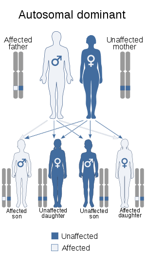 | |
| This condition is inherited in an autosomal dominant manner | |
| Specialty | Ophthalmology |
| Symptoms | Lattice degeneration itself does not cause symptoms |
| Diagnostic method | The only way to diagnose the condition is with a dilated fundus examination by an eye care provider. A dilated fundus examination is done by administering dilating eye drops in your eyes to expand the pupil so that the retina can be carefully evaluated. Dilating drops will cause your vision to be blurry for several hours before returning to normal. |
Lattice degeneration occurs in approximately 6–8% of the general population and in approximately 30% of phakic retinal detachments.[1] Similar lesions are seen in patients with Ehlers-Danlos syndrome, Marfan syndrome, and Stickler syndrome, all of which are associated with an increased risk of retinal detachment. Risk of developing lattice degeneration in one eye is also increased if lattice degeneration is already present in the other eye.[1]
Signs
Typical lattice consists of sharply demarcated, spindle-shaped areas of retinal thinning, usually located between the equator of the retina and the posterior border of the vitreous base. This is more frequently located in the temporal half of the retina and is seen more superiorly than inferiorly.
Atypical lattice is characterised by radial lesions which appear continuous with the peripheral blood vessels. This type is typically seen in patients with Stickler syndrome.
Diagnosis
Treatment
Barrage laser is at times done prophylactically around a hole or tear associated with lattice degeneration in an eye at risk of developing a retinal detachment. It is not known if surgical interventions such as laser photocoagulation or cryotherapy is effective in preventing retinal detachment in patients with lattice degeneration or asymptomatic retinal detachment.[1] Laser photocoagulation has been shown to reduce risks of retinal detachment in symptomatic lattice degeneration. There are documented cases wherein retina detached from areas which were otherwise healthy despite being treated previously with laser.
Prognosis
No complications are encountered in most patients with lattice degeneration, although in young myopes, retinal detachment can occur. There are documented cases with macula-off retinal detachment in patients with asymptomatic lattice degeneration. Partial or complete vision loss almost always occurs in such cases. Currently there is no prevention or cure for lattice degeneration.
References
- Wilkinson, Charles P. (2014-09-05). "Interventions for asymptomatic retinal breaks and lattice degeneration for preventing retinal detachment". The Cochrane Database of Systematic Reviews (9): CD003170. doi:10.1002/14651858.CD003170.pub4. ISSN 1469-493X. PMC 4423540. PMID 25191970.