Radiographic classification of osteoarthritis
Radiographic systems to classify osteoarthritis vary by which joint is being investigated. In osteoarthritis, the choice of treatment is based on pain and decreased function, but radiography can be useful before surgery in order to prepare for the procedure.
| Radiographic classification of osteoarthritis | |
|---|---|
| Medical diagnostics | |
| Purpose | Quantify the degree of osteoarthritis |
| Based on | Medical imaging |
Vertebral column
There are many grading systems for degeneration of intervertebral discs and facet joints in the cervical and lumbar vertebrae, of which the following radiographic systems can be recommended in terms of interobserver reliability:[1]
- Kellgren grading of cervical disc degeneration
- Kellgren grading of cervical facet joint degeneration
- Lane grading of lumbar disc degeneration
- Thompson grading of lumbar disc degeneration (by Magnetic resonance imaging)
- Pathria grading of lumbar facet joint degeneration (by computed tomography)
- Weishaupt grading of lumbar facet joint degeneration (by MRI and computed tomography)
| I |
|
|---|---|
| II |
|
| III |
|
| IV |
|
| Grade | Joint space narrowing | Osteophytes | Sclerosis |
|---|---|---|---|
| 0 | None | None | None |
| 1 | Definite but mild narrowing | Small | Present |
| 2 | Moderate | Moderate | – |
| 3 | Severe (complete joint space loss) | Large | – |
The Thomson grading system is regarded to have more academic than clinical value.[1]
| Grade | Nucleus | Anulus | Endplate | Vertebral body |
|---|---|---|---|---|
| I | Bulging gel | Discrete fibrous laminae | Hyaline, uniform thickness | Rounded margins |
| II | Peripheral white fibrous tissue | Mucinous material between laminae | Irregular thickness | Pointed margins |
| III | Consolidated fibrous tissue | Extensive mucinous infiltration; loss of annular-nuclear demarcation | Focal defects in cartilage | Small chondrophytes or osteophytes at margins |
| IV | Horizontal clefts parallel to endplate | Focal disruptions | Fibrocartilage extending from subchondral bone; irregularity and focal sclerosis in subchondral bone | Osteophytes smaller than 2 mm |
| V | Clefts extended through nucleus and annulus | Diffuse sclerosis | Osteophytes greater than 2 mm |
Shoulder
The Samilson-Prieto classification is preferable for osteoarthritis of the glenohumeral joint.[3]
| Grade | Description |
|---|---|
| Mild | Exostosis of inferior humerus and/or glenoid measuring less than 3 mm |
| Moderate | Exostosis of inferior humerus and/or glenoid measuring 3–7 mm, and slight irregularity of the joint |
| Severe | Exostosis of inferior humerus and/or glenoid measuring more than 7 mm in height as well as sclerosis and narrowing of the joint space (normal joint space is 4–5 mm).[5] |
Hip
.jpg)
The most commonly used radiographic classification system for osteoarthritis of the hip joint is the Kellgren-Lawrence system (or K-L system).[6] It uses plain radiographs.
| Grade | Description |
|---|---|
| 0 | No radiographic features of osteoarthritis |
| 1 | Possible joint space narrowing (normal joint space is at least 2 mm at the superior acetabulum)[7] and osteophyte formation |
| 2 | Definite osteophyte formation with possible joint space narrowing |
| 3 | Multiple osteophytes, definite joint space narrowing, sclerosis and possible bony deformity |
| 4 | Large osteophytes, marked joint space narrowing, severe sclerosis and definite bony deformity |
Osteoarthritis of the hip joint may also be graded by Tönnis classification. There is no consensus whether it is more or less reliable than the Kellgren-Lawrence system.[8]
_osteoarthritis_of_the_hip.jpg)
| Grade | Description |
|---|---|
| 0 | No osteoarthritis signs |
| 1 | Mild:
|
| 2 | Moderate:
|
| 3 | Severe:
|
Knee
For the grading of osteoarthritis in the knee, the International Knee Documentation Committee (IKDC) system is regarded to have the most favorable combination of interobserver precision and correlation to knee arthroscopy findings.[10] It was formed by a group of knee surgeons from Europe and America who met in 1987 to develop a standard form to measure results of knee ligament reconstructions.[11]
The Ahlbäck system has been found to have comparable interobserver precision and arthroscopy correlation to the IKDC system, but most of the span of the Ahlbäck system focused at various degrees of bone defect or loss, and it is therefore less useful in early osteoarthritis.[10] Systems that have been found to have lower interobserver precision and/or arthroscopy correlation are those developed by Kellgren-Lawrence, Fairbank, Brandt, and Jäger-Wirth.[10]
| Grade | Findings |
|---|---|
| A | No joint space narrowing, defined in this system as at least 4 mm joint space |
| B | At least 4 mm joint space, but small osteophytes, slight sclerosis, or femoral condyle flattening |
| C | 2–4 mm joint space |
| D | <2 mm joint space |
| Grade | Findings |
|---|---|
| I | Joint space narrowing, with or without subchondral sclerosis. Joint space narrowing is defined by this system as a joint space less than 3 mm, or less than half of the space in the other compartment, or less than half of the space of the homologous compartment of the other knee. |
| II | Obliteration of the joint space |
| III | Bone defect/loss <5 mm |
| IV | Bone defect/loss between 5 and 10 mm |
| V | Bone defect/loss >10 mm, often with subluxation and arthritis of the other compartment |
For the patellofemoral joint, a classification by Merchant 1974 uses a 45° "skyline" view of the patella:[13]
| Stage | Description |
|---|---|
| 1 (mild) | Patellofemoral joint space > 3mm |
| 2 (moderate | Joint space < 3 mm but no bony contact |
| 3 (severe) | Bony surfaces in contact over less than one quarter of the joint surface |
| 4 (very severe) | Bony contact throughout the entire joint surface |
Other joints
- In the temporomandibular joint, subchondral sclerosis of the mandibular condyle has been described as an early change, condylar flattening as a feature of progressive osteoarthritis, and narrowing of the temporomandibular joint space as a late stage change.[14] A joint space of between 1.5 and 4 mm is regarded as normal.[15]
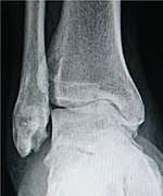
- For the ankle, the Kellgren-Lawrence scale, as described for the hip, has been recommended.[16] The distances between the bones in the ankle are normally as follows:[17]
- Talus - medial malleolus : 1.70 ± 0.13 mm
- Talus - tibial plafond: 2.04 ± 0.29 mm
- Talus - lateral malleolus: 2.13 ± 0.20 mm
See also
References
- Kettler, Annette; Wilke, Hans-Joachim (2005). "Review of existing grading systems for cervical or lumbar disc and facet joint degeneration". European Spine Journal. 15 (6): 705–718. doi:10.1007/s00586-005-0954-y. ISSN 0940-6719. PMC 3489462.
- Ofiram, Elisha; Garvey, Timothy A.; Schwender, James D.; Denis, Francis; Perra, Joseph H.; Transfeldt, Ensor E.; Winter, Robert B.; Wroblewski, Jill M. (2009). "Cervical degenerative index: a new quantitative radiographic scoring system for cervical spondylosis with interobserver and intraobserver reliability testing". Journal of Orthopaedics and Traumatology. 10 (1): 21–26. doi:10.1007/s10195-008-0041-3. ISSN 1590-9921. PMC 2657349.
- Brox, Jens; Lereim, Paul; Merckoll, Else; Finnanger, Anne Marie (2009). "Radiographic classification of glenohumeral arthrosis". Acta Orthopaedica Scandinavica. 74 (2): 186–189. doi:10.1080/00016470310013932. ISSN 0001-6470.
- Page 117 in Barbara N. W. Weissman (2009). Imaging of Arthritis and Metabolic Bone Disease. Elsevier Health Sciences. ISBN 9780323041775.
- "Glenohumeral joint space". radref.org., in turn citing: Petersson, Claes J.; Redlund-Johnell, Inga (2009). "Joint Space in Normal Gleno-Humeral Radiographs". Acta Orthopaedica Scandinavica. 54 (2): 274–276. doi:10.3109/17453678308996569. ISSN 0001-6470.
- Zdravko Jotanovic, Radovan Mihelic, Gordan Gulan, Branko Sestan, Zlatko Dembic (2015). "Osteoarthritis of the hip: An overview". Periodicum biologorum. 117 (1).CS1 maint: multiple names: authors list (link)
- Lequesne, M (2004). "The normal hip joint space: variations in width, shape, and architecture on 223 pelvic radiographs". Annals of the Rheumatic Diseases. 63 (9): 1145–1151. doi:10.1136/ard.2003.018424. ISSN 0003-4967. PMC 1755132.
- Terjesen, Terje; Gunderson, Ragnhild B (2012). "Radiographic evaluation of osteoarthritis of the hip". Acta Orthopaedica. 83 (2): 185–189. doi:10.3109/17453674.2012.665331. ISSN 1745-3674. PMC 3339535.
- "Tönnis Classification of Osteoarthritis by Radiographic Changes". Society of Preventive Hip Surgery. Retrieved 2016-12-13.
- "Osteoarthritis Classification Scales: Interobserver Reliability and Arthroscopic Correlation". The Journal of Bone and Joint Surgery. American Volume. 96 (14): 1145–1151. 2014. doi:10.2106/JBJS.M.00929. ISSN 0021-9355. PMC 4083772.
- Hefti F, Müller W, Jakob RP, Stäubli HU (1993). "Evaluation of knee ligament injuries with the IKDC form". Knee Surg Sports Traumatol Arthrosc. 1 (3–4): 226–34. doi:10.1007/bf01560215. PMID 8536037.
- Hernández-Vaquero, Daniel; Fernández-Carreira, José Manuel (2012). "Relationship between radiological grading and clinical status in knee osteoarthritis. a multicentric study". BMC Musculoskeletal Disorders. 13 (1). doi:10.1186/1471-2474-13-194. ISSN 1471-2474.
- Kim, Young-Mo; Joo, Yong-Bum (2012). "Patellofemoral Osteoarthritis". Knee Surgery & Related Research. 24 (4): 193–200. doi:10.5792/ksrr.2012.24.4.193. ISSN 2234-0726. PMC 3526755.
- Page 722 in Gary S. Firestein, Ralph Budd, Sherine E Gabriel, Iain B. McInnes, James R O'Dell (2012). Kelley's Textbook of Rheumatology E-Book. Elsevier Health Sciences. ISBN 9781455737673.CS1 maint: multiple names: authors list (link)
- Massilla Mani, F.; Sivasubramanian, S. Satha (2016). "A study of temporomandibular joint osteoarthritis using computed tomographic imaging". Biomedical Journal. 39 (3): 201–206. doi:10.1016/j.bj.2016.06.003. ISSN 2319-4170.
- Nicolas Holzer, Davide Salvo, Anne Karien Marijnissen, Aminudin Che Ahmad, Emanuele Sera, Pierre Hoffmeyer, Anne Lübbeke Wolff, Mathieu Assal (2017-09-14). "How to assess ankle osteoarthritis: comparison of the Kellgren and Lawrence scale with functional outcome and digital image analysis". Orthopaedic Proceedings. 94-B.CS1 maint: multiple names: authors list (link)
- Imai, Kan; Ikoma, Kazuya; Kido, Masamitsu; Maki, Masahiro; Fujiwara, Hiroyoshi; Arai, Yuji; Oda, Ryo; Tokunaga, Daisaku; Inoue, Nozomu; Kubo, Toshikazu (2015). "Joint space width of the tibiotalar joint in the healthy foot". Journal of Foot and Ankle Research. 8 (1). doi:10.1186/s13047-015-0086-5. ISSN 1757-1146.
- Quintana, José M.; Escobar, Antonio; Arostegui, Inmaculada; Bilbao, Amaia; Azkarate, Jesús; Goenaga, J. Ignacio; Arenaza, Juan C. (2006). "Health-Related Quality of Life and Appropriateness of Knee or Hip Joint Replacement". Archives of Internal Medicine. 166 (2): 220. doi:10.1001/archinte.166.2.220. ISSN 0003-9926.