Intraepithelial lymphocyte
Intraepithelial lymphocytes (IEL) are lymphocytes found in the epithelial layer of mammalian mucosal linings, such as the gastrointestinal (GI) tract and reproductive tract.[1] However, unlike other T cells, IELs do not need priming. Upon encountering antigens, they immediately release cytokines and cause killing of infected target cells. In the GI tract, they are components of gut-associated lymphoid tissue (GALT).[2]
Based on expression of either an αβ T-cell receptor (TCR) or a γδ TCR IEL T cells can be divide into two major groups. In mice both groups are retained in almost equal proportions.[3] In humans, the majority of IELs are alpha beta T cells. 15% of IELs are gamma delta T cells and thus represent a minor component of human IELs. However, IELs significantly increase under certain conditions, such as celiac disease.[1]
Phenotype
The majority of IELs (80%) are CD3+, and over 75% of these also express CD8. IELs can be divide into two major subsets based on their CD8 coreceptor expression.[3] One subset of IELs typically express activation marker CD8αα and some IELs express CD8αβ+ marker (CD8αβ promotes TCR activation, whereas CD8αα suppresses TCR signals).
In both humans and mice IELs express higher levels of CD103, activation marker CD69, granzyme B and perforin cytolytic granules. CD25 expression is lower in comparison with effector memory T cells.[4][5]
Development
Induced IELs (TCRαβ+ CD8αβ+) are generated from naive T cells during an immune response. TCRαβ+ CD8αα (natural IELs) cells differentiate in the thymus.[4][6]
Development and cytolytic activation are independent of live micro-organisms but they become cytolytic in response to the exogenous antigenic substances other than live micro-organisms in the gut. EIL T cells acquired their activated memory phenotype post-thymically, in response to antigens encountered in the periphery.[7]
Function
Their role in immune system is crucial because IELs provide a first line of defense at this extensive barrier with the outside world. All IEL T cells are antigen-experienced T cells, which typically display a cytotoxic functional phenotype. IELs mediate antigen-specific delayed-type hypersensitivity (DTH) responses, exhibit virus-specific CTL function, to express natural killer (NK)-like activity and produce a local graft-versus-host reaction (GVHR) when transferred to semiallogeneic hosts. IELs are also able to produce a variety of cytokines which are characteristically produced by Th1- and Th2-type cells and can also provide help for B cell responses.[4][6][7]
Pathology
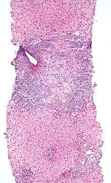
An elevated IEL population, as determined by biopsy, typically indicates ongoing inflammation within the mucosa. In diseases such as celiac sprue, IEL elevation throughout the small intestine is one of many specific markers.[1] IELs have heightened activated status that can lead to inflammatory disease such as IBD, promote cancer development and progression,[8] or become the malignant cells in enteropathy-associated T-cell lymphoma, a lymphoma that is a complication of celiac sprue.[9][10]
Alternatively, elevated IEL populations can be a marker for developing neoplasia in the tissue such as found in cervical and prostate cancers, as well as some colorectal cancers, particularly those associated with Lynch syndrome (hereditary non-polyposis colon cancer <HNPCC>).[11] IELs themselves can, when chronically activated, undergo mutation that can lead to lymphoma.[12]
See also
References
- Hopper AD, Hurlstone DP, Leeds JS, McAlindon ME, Dube AK, Stephenson TJ, Sanders DS (November 2006). "The occurrence of terminal ileal histological abnormalities in patients with coeliac disease". Digestive and Liver Disease. 38 (11): 815–9. doi:10.1016/j.dld.2006.04.003. PMID 16787773.
- Defranco, Anthony L; Locksley, Richard M; Robertson, Miranda. 2007. Immunity: The Immune Response in Infection and Inflammatory Disease. New Science Press Ltd. 218-219.
- Sheridan BS, Lefrançois L (December 2010). "Intraepithelial lymphocytes: to serve and protect". Current Gastroenterology Reports. 12 (6): 513–21. doi:10.1007/s11894-010-0148-6. PMC 3224371. PMID 20890736.
- Mayassi T, Jabri B (September 2018). "Human intraepithelial lymphocytes". Mucosal Immunology. 11 (5): 1281–1289. doi:10.1038/s41385-018-0016-5. PMC 6178824. PMID 29674648.
- Lambolez F, Mayans S, Cheroutre H (2013). Lymphocytes: Intraepithelial. eLS. American Cancer Society. doi:10.1002/9780470015902.a0001197.pub3. ISBN 9780470015902.
- Sim GK (1995-01-01). "Intraepithelial lymphocytes and the immune system". Advances in Immunology. 58: 297–343. doi:10.1016/s0065-2776(08)60622-7. ISBN 9780120224586. PMID 7741030.
- McGhee, Jerry R. (1998-01-01). Delves, Peter J. (ed.). "Mucosa-Associated Lymphoid Tissue (MALT)". Encyclopedia of Immunology (Second ed.). Elsevier: 1774–1780. doi:10.1006/rwei.1999.0448. ISBN 9780122267659.
- Cheroutre H, Lefrancois L (2015-01-01). "Chapter 35 - Intraepithelial TCRαβ T Cells in Health and Disease". In Mestecky J, Strober W, Russell MW, Kelsall BL (eds.). Mucosal Immunology (Fourth ed.). Academic Press. pp. 733–748. doi:10.1016/b978-0-12-415847-4.00035-5. ISBN 9780124158474.
- Ondrejka S, Jagadeesh D (December 2016). "Enteropathy-Associated T-Cell Lymphoma". Current Hematologic Malignancy Reports. 11 (6): 504–513. doi:10.1007/s11899-016-0357-7. PMID 27900603.
- Chander U, Leeman-Neill RJ, Bhagat G (August 2018). "Pathogenesis of Enteropathy-Associated T Cell Lymphoma". Current Hematologic Malignancy Reports. 13 (4): 308–317. doi:10.1007/s11899-018-0459-5. PMID 29943210.
- Bellizzi AM, Frankel WL (November 2009). "Colorectal cancer due to deficiency in DNA mismatch repair function: a review". Advances in Anatomic Pathology. 16 (6): 405–17. doi:10.1097/PAP.0b013e3181bb6bdc. PMID 19851131.
- Meresse B, Malamut G, Cerf-Bensussan N (June 2012). "Celiac disease: an immunological jigsaw". Immunity. 36 (6): 907–19. doi:10.1016/j.immuni.2012.06.006. PMID 22749351.