Internal vertebral venous plexuses
The internal vertebral venous plexuses (intraspinal veins) lie within the vertebral canal in the epidural space,[1] and receive tributaries from the bones and from the spinal cord.
| Internal vertebral venous plexuses | |
|---|---|
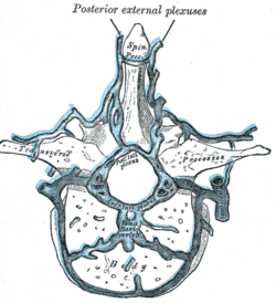 Transverse section of a thoracic vertebra, showing the vertebral venous plexuses. | |
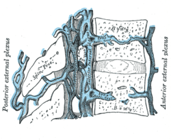 Median sagittal section of two thoracic vertebrae, showing the vertebral venous plexuses. | |
| Details | |
| Identifiers | |
| Latin | plexus venosus vertebralis internus |
| TA | A12.3.07.021 A12.3.07.026 |
| FMA | 4881 |
| Anatomical terminology | |
They form a closer network than the external plexuses, and, running mainly in a vertical direction, form four longitudinal veins, two in front and two behind; they therefore may be divided into anterior and posterior groups.
- The anterior internal plexuses consist of large veins which lie on the posterior surfaces of the vertebral bodies and intervertebral fibrocartilages on either side of the posterior longitudinal ligament; under cover of this ligament they are connected by transverse branches into which the basivertebral veins open.
- The posterior internal plexuses are placed, one on either side of the middle line in front of the vertebral arches and ligamenta flava, and anastomose by veins passing through those ligaments with the posterior external plexuses.
The anterior and posterior plexuses communicate freely with one another by a series of venous rings (retia venosa vertebrarum), one opposite each vertebra.
Around the foramen magnum they form an intricate network which opens into the vertebral veins and is connected above with the occipital sinus, the basilar plexus, the condyloid emissary vein, and the rete canalis hypoglossi.
References
This article incorporates text in the public domain from page 668 of the 20th edition of Gray's Anatomy (1918)
- Moore, Keith (2010). Clinically Oriented Anatomy. Philadelphia: Lippincott Williams and Wilkins. pp. 472–3. ISBN 978-0-7817-7525-0.
External links
- Atlas image: abdo_wall77 at the University of Michigan Health System - "Venous Drainage of the Vertebral Column"
| Authority control |
|---|