Internal thoracic artery
In human anatomy, the internal thoracic artery (ITA), previously known as the internal mammary artery (a name still common among surgeons), is an artery that supplies the anterior chest wall and the breasts. It is a paired artery, with one running along each side of the sternum, to continue after its bifurcation as the superior epigastric and musculophrenic arteries.
| Internal thoracic artery | |
|---|---|
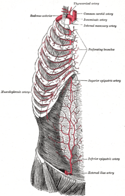 Right internal thoracic artery and its branches (labeled under its old name the Internal mammary artery, at upper right.) | |
| Details | |
| Source | Subclavian artery |
| Branches | Pericardiocophrenic Anterior intercostal branches Musculophrenic Superior epigastric Perforating branches |
| Vein | Internal thoracic vein |
| Identifiers | |
| Latin | Arteria thoracica interna, arteria mammaria interna |
| MeSH | D008323 |
| TA | A12.2.08.029 |
| FMA | 3960 |
| Anatomical terminology | |
Structure
The internal thoracic artery arises from the subclavian artery near its origin.
It travels downward on the inside of the ribcage, approximately a centimeter from the sides of the sternum, and thus medial to the nipple. It is accompanied by the internal thoracic vein.
It runs deep to the external oblique, but superficial to the vagus nerve
Branches
- Mediastinal branches
- Thymic branches
- Pericardiacophrenic artery - travels with the phrenic nerve
- Sternal branches
- Perforating branches
- Twelve anterior intercostal branches, two to each of the top six intercostal spaces. In a given space, the upper branch travelling laterally along the bottom of the rib until it anastomoses with its corresponding posterior intercostal artery. The lower branch of the space anastomoses with a collateral branch of the posterior intercostal artery.
After passing the sixth intercostal space, the internal thoracic artery splits into the following two terminal branches:
- Musculophrenic artery - roughly follows the costal margin
- Superior epigastric artery - continues the course of the internal thoracic artery, travelling downward into the abdominal wall
Clinical significance
Use in bypass grafts
The internal thoracic artery is the cardiac surgeon's blood vessel of choice for coronary artery bypass grafting. The left ITA has a superior long-term patency to saphenous vein grafts[1][2] and other arterial grafts[3] (e.g. radial artery, gastroepiploic artery) when grafted to the left anterior descending coronary artery, generally the most important vessel, clinically, to revascularize.
Plastic surgeons may use either the left or right internal thoracic arteries for autologous free flap reconstruction of the breast after mastectomy. Usually, a microvascular anastomosis is performed at the second intercostal space to the artery on which the free flap is based.
Additional images
 Anterior Thoracic Wall, from behind
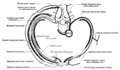 Diagram of an Intercostal Space
References
- Kitamura, S; Kawachi, K; Kawata, T; Kobayashi, S; Mizuguchi, K; Kameda, Y; Nishioka, H; Hamada, Y; Yoshida, Y (1996). "Ten-year survival and cardiac event-free rates in Japanese patients with the left anterior descending artery revascularized with internal thoracic artery or saphenous vein graft: a comparative study". Nippon Geka Gakkai Zasshi. 97 (3): 202–9. PMID 8649330.
- Arima, M; Kanoh, T; Suzuki, T; Kuremoto, K; Tanimoto, K; Oigawa, T; Matsuda, S (2005). "Serial angiographic follow-up beyond 10 years after coronary artery bypass grafting". Circulation Journal. 69 (8): 896–902. doi:10.1253/circj.69.896. PMID 16041156.
- Cohen, G; Tamariz, MG; Sever, JY; Liaghati, N; Guru, V; Christakis, GT; Bhatnagar, G; Cutrara, C; et al. (2001). "The radial artery versus the saphenous vein graft in contemporary CABG: a case-matched study". The Annals of Thoracic Surgery. 71 (1): 180–5, discussion 185–6. doi:10.1016/S0003-4975(00)02285-2. PMID 11216742.
External links
| Wikimedia Commons has media related to Internal thoracic artery. |
Figures of ITA grafts
- Figure of heart with two saphenous vein grafts (SVGs) and a LITA graft - texheartsurgeons.com
- Drawing of the heart with a SVG to the right coronary artery (RCA) and a LITA graft to the LAD - darcystudios.com
- Drawing of the heart with a SVG to the RCA and a LITA graft to the LAD - mayoclinic.org
| Authority control |
|---|