Instruments used in radiology
Instruments used specially in radiology are as follows:[1][2][3]
| Instrument | Uses |
|---|---|
| Ultrasonography machine | uses ultrasound to produce images from within the body; video link |
| X-ray | uses X-rays to produce images of structures within the body; video link |
| Contrast media for X-rays | to provide a high contrast image of the details of the viscera under study; e.g. salts of heavy metals, gas like air, radio-opaque dyes, organic iodides, etc. |
| Echocardiography machine | sonography of the heart is done here to know its function and state |
| Computer axial tomography scan (CT Scan)/(CAT Scan) | to visualize the interior of the body in slices (traditionally showing horizontal slices); video link |
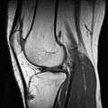
| Magnetic resonance imaging (MRI) alias Nuclear magnetic resonance (NMR) | high strength (0.15 to 1.5 teslas)[4] are used to excite protons that produce the record results (like CT scan). It can show particular tissues more clearly than CT.;[4] video link |
| Linear accelerator | used in radiotherapy for cancer |
| Functional magnetic resonance imaging (fMRI) | video link |
| Positron emission tomography (PET Scan) | video link |
| Radio-isotope scan or nuclear scintigraphy | These radioactive compounds are administered so that specific tissues take them up. The amount and anatomical detail of the uptake produces the scan result. |
| SPECT scan | video link |
| Interventional radiology | minimally invasive surgeries under radiological imaging, e.g. angioplasty, TIPS. |
| Brachytherapy apparatus | video link |
| Lead shielding | visual and physical protection from x-ray |
Image gallery
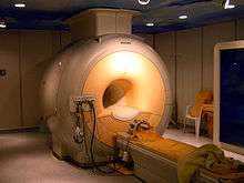 A 3 tesla MRI scanner
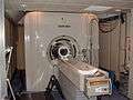 fMRI scanner
 Rotating anode X-ray tube
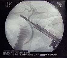 X-ray guided cholecystectomy
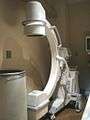 Mobile fluoroscopy machine
References
- Arun Baran Singha Mahapatra. Essentials of medical physiology. ISBN 81-86793-56-9.
- P. Chakraborty; G. Chakraborty. Practical Pathology. ISBN 81-7381-332-9.
- Robbins and Cotran Review of Pathology. ISBN 0-7216-0194-4.
- David Sutton. Radiology and imaging for med. students (7th ed.). ISBN 81-7867-100-X.
This article is issued from
Wikipedia.
The text is licensed under Creative
Commons - Attribution - Sharealike.
Additional terms may apply for the media files.