Port (medical)
In medicine, a port is a small medical appliance that is installed beneath the skin. A catheter connects the port to a vein. Under the skin, the port has a septum through which drugs can be injected and blood samples can be drawn many times, usually with less discomfort for the patient than a more typical "needle stick".
Ports are used mostly to treat hematology and oncology patients. Ports were previously adapted for use in hemodialysis patients, but were found to be associated with increased rate of infections and are no longer available in the US.[1]
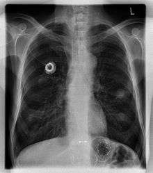
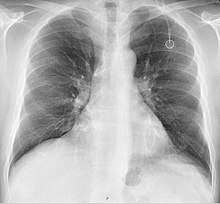
The port is usually inserted in the upper chest (known as a "chest port"), just below the clavicle or collar bone, leaving the patient's hands free.
Terminology
A port is more correctly known as a "totally implantable venous access device". Brand Names include Eco Port, Clip-a-Port, SmartPort, Microport, Bardport, PowerPort, Passport, Port-a-Cath, Infuse-a-Port, Medi-Port, and Bioflo.
How it works
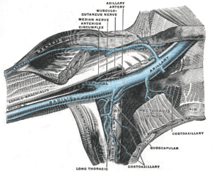
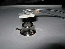
A port consists of a reservoir compartment (the portal) that has a silicone bubble for needle insertion (the septum), with an attached plastic tube (the catheter). The device is surgically inserted under the skin in the upper chest or in the arm and appears as a bump under the skin. It requires no special maintenance other than occasional flushing to keep clear. It is completely internal so swimming and bathing are not a problem. The catheter runs from the portal and is surgically inserted into a vein (usually the jugular vein or less optimally the subclavian vein). Ideally, the catheter terminates in the superior vena cava or the right atrium. This position allows infused agents to be spread throughout the body quickly and efficiently.
The septum is made of a special self-sealing silicone; it can be punctured hundreds of times before it weakens significantly. To administer treatment or to withdraw blood, a health care professional will first locate the port and disinfect the area, then access the port by puncturing the overlying skin with a Huber point (non-coring) needle. Due to its design, there is a very low infection risk, as the breach of skin integrity is never larger than the caliber of the needle. This gives it an advantage over indwelling lines such as the Hickman line. Negative pressure is created to withdraw blood into the vacuumized needle, to check for blood return and see if the port is functioning normally. Next, the port is flushed with a saline solution. Then, treatment will begin.
The implantation procedure itself is considered minor, and is typically performed with both local anaesthesia and moderate sedation. Patients often have post-procedure discomfort at the insertion site which is most often managed by administration of acetaminophen or a non-steroidal anti-inflammatory drug such as ibuprofen.
A port is most commonly inserted as an outpatient surgery procedure in a hospital or clinic by an interventional radiologist or surgeon, under moderate sedation. Implantation is increasingly performed by interventional radiologists due to advancements in techniques and their facile use of imaging technologies. When no longer needed, the port can be removed in the interventional radiology suite or an operating room.
Uses
Ports have many uses:
- To deliver chemotherapy to cancer patients who must undergo treatment frequently. Chemotherapy is often toxic, and can damage skin and muscle tissue, and therefore should not be delivered through these tissues. Ports provide a solution, delivering drugs quickly and efficiently through the entire body via the circulatory system.
- To deliver coagulation factors in patients with severe hemophilia.
- To withdraw (and/or return) blood to the body in patients who require frequent blood tests, and in hemodialysis patients.
- To deliver antibiotics to patients requiring them for a long time or frequently, such as those with cystic fibrosis and bronchiectasis.
- Delivering medications to patients with immune disorders.
- For treating alpha 1-antitrypsin deficiency with replacement therapy
- For delivering radiopaque contrast agents, which enhance contrast in CT imaging.
- To fill or withdraw fluid from the Lap-Band or Realize gastric bands used in Bariatric surgeries.
- To administer analgesics to patients with chronic pain, such as cancer patients and those with sickle-cell disease
Insertion
Fluoroscopy is useful in guiding the insertion of ports.[2]
A follow-up chest radiograph can immediately detect complications associated with the procedure in the form of pneumothorax, hemothorax and malpositions of the catheter (see Risks below for further details). However, it is suggested that chest radiography is not mandatory as a routine method after fluoroscopy-guided port insertion that is mainly performed by venous cutdown.[2]
The side of the patients' chest the port is implanted in will usually be chosen to avoid damage to the port and the veins by the seat belt in case of accident when seated as the driver. Thus, there is a potential conflict by left- and right-hand traffic as the rule of the road.[3][4]
Models
There are many different models of ports. The particular model selected is based on the patient's specific medical conditions.
Portals:
- can be made of plastic, stainless steel, or titanium
- can be single chamber or dual chamber
- vary in height, width and shape.
Catheters:
- can be made of biocompatible, medical-grade polyurethane or silicone
- can vary in length and diameter
Ports can be put in the upper chest or arm. The exact positioning itself is variable as it can be inserted to avoid visibility when wearing low cut shirts, and to avoid excess contact due to a backpack or bra strap. The most common placement is on the upper right portion of the chest, with the catheter itself looping through the right jugular vein, and down towards the patient's heart.
For applications as CT scan, high pressure infusion allowing ports are needed.[5][6]
Risks
- Age: If the device is put into a child, the child's growth means that the catheter becomes relatively shorter and will move towards the head. It may become necessary to remove or replace it.
- Arterial injury: The subclavian artery can be inadvertently punctured while attempting a subclavian vein access, leading to a subcutaneous hematoma and occasionally a pseudoaneurysm. An alternative site may need to be used for port placement. Puncture of the carotid artery is significantly more rare, since attempts to access the nearby jugular vein are increasingly done with ultrasound guidance.
- Infection: An infection may develop in the line or around the port. This may require antibiotic treatment or removal of the device.
- Mechanical failure is uncommon. Ports placed through the subclavian vein may suffer from "pinch-off syndrome" where the catheter fractures as it passes into the vein. Ports placed via the jugular vein do not suffer from this problem. The catheter fragment then travels through the venous system and typically lodges in the right heart or the lungs. Many patients are asymptomatic but the mechanical failure is discovered because of an inability to flush or withdraw fluids from the port. In those instances, an interventional radiologist can usually retrieve the fragment and place a new port.
- Pneumothorax: Attempts to gain access to the subclavian vein or jugular vein can injure the lung, potentially causing a pneumothorax. If the pneumothorax is large enough, a chest tube might need to be placed. In experienced hands, the incidence of this complication is about 1% when accessing the subclavian vein. When accessing the jugular vein the pneumothorax rate is virtually nonexistent.
- Thrombosis: formation of a blood clot in the catheter may block the device irrevocably. To prevent clotting the port is flushed with saline and heparin, usually by a nurse or other medical professional, or someone properly trained that is a family member or the patient, once every six weeks, or more often in conjunction with administering medication.
- Vascular occlusion: formation of a blood clot between the catheter and the vascular wall leading to partial or complete occlusion of the vein. The occlusion is cleared by removal of the port if possible. If not, then heparin therapy may clear the occlusion.
Manufacturers
The major manufacturers of ports are AngioDynamics, B. Braun Medical,[7] Bard Access Systems,[6] Cook Medical, MedComp, Navilyst Medical, Norfolk Medical Products, and Smiths Medical.
Use
To reduce damage or coring of the septum during use, low or non coring needles are to be used.[8] After each use, a heparin lock is made by injecting a small amount of heparinized saline (an anticoagulant) into the device, preventing development of clots within the port or catheter. In some catheter designs where there is a self-sealing valve at the far end, the system is locked with just saline. The port can be left accessed for as long as required. The port is covered in a dressing to protect the site from infection and to secure the needle in position.
If a port is used infrequently, it may be necessary to access the port, flush it with saline, and inject a new heparin lock to prevent clotting between uses.
Alternatives
Sometimes, the physical condition of the patient, especially the structure of his veins, does not allow for the insertion of a port. An alternative is the PICC line, despite drawbacks such as external entry point and limited lifespan of the device.[9]
In popular culture
In the 1984 cyberpunk novel Neuromancer, a minor character, Peter Riviera, has a kind of medical port placed in his arm to facilitate his recreational drug use.[10]
See also
References
- "Gastroenterology-Urology Devices; Reclassification of Implanted Blood Access Devices". Food and Drug Administration. 25 July 2014.
- Thomopoulos, Theodoros; Meyer, Jeremy; Staszewicz, Wojciech; Bagetakos, Ilias; Scheffler, Max; Lomessy, Antoine; Toso, Christian; Becker, Christoph D.; Morel, Philippe (2014). "Routine Chest X-ray is not Mandatory after Fluoroscopy-Guided Totally Implantable Venous Access Device Insertion". Annals of Vascular Surgery. 28 (2): 345–350. doi:10.1016/j.avsg.2013.08.003. ISSN 0890-5096.
- Julia Lederbogen-Hülsen (2009). Erleichterung der Chemotherapie durch implantierbare Portkatheter-Systeme bei Patientinnen mit gynäkologischen Tumoren (in German). Münster: Universitätsklinikum Münster. p. 91.
Verlauf des Autosicherheitsgurts in die Überlegungen mit einzubeziehen (to include the place of the safety belt into the planning)
- "Celsite® Portkatheter-Systeme" (PDF) (in German). B. Braun Melsungen. 2012.
Auf welcher Seite wird der Sicherheitsgurt angebracht? (which side is the safety belt)
- "C-Port®CT". Retrieved 25 November 2017.
- "IMPLANTABLE PORT DEVICES". Retrieved 23 November 2017.
- "Celsite® Access Ports" (PDF). Retrieved 23 November 2017.
- "Choice of the Needles" (PDF). p. 7. Retrieved 25 November 2017.
- Michaela Hans. "Pflegeleitfaden" (PDF) (in German). CHARITÉ. p. 22. Retrieved 2017-12-03.
Liegedauer von 4 Monaten
- Gibson, William (July 2000) [July 1983]. "Chapter Eight". Neuromancer (Ace trade paperback ed.). p. 105.
Riviera loosened and removed the elastic length of surgical tubing from his arm. 'Yes. It's more fun.' He smiled, his eyes distant now, cheeks flushed. 'I've a membrane set in, just over the vein, so I never have to worry about the condition of the needle.' 'Doesn't hurt?' [said Case] The bright eyes met his. 'Of course it does. That's part of it, isn't it?'
Further reading
- Mallon, William (March 2001). "Is It Acceptable to Discharge a Heroin User with an Intravenous Line to Complete His Antibiotic Therapy for Cellulitis at Home under a Nurse's Supervision?". Point-Counterpoint (column). The Western Journal of Medicine. 174 (3): 157. doi:10.1136/ewjm.174.3.157. PMC 1071292. PMID 11238332.
External links
| Wikimedia Commons has media related to Implantable venous access devices. |