Hypertrophic decidual vasculopathy
In pathology, hypertrophic decidual vasculopathy, abbreviated HDV, is the histomorphologic correlate of gestational hypertension, as may be seen in intrauterine growth restriction (IUGR)[1] and HELLP syndrome.
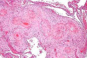
Micrograph showing hypertrophic decidual vasculopathy, the histomorphologic correlate of gestational hypertension. H&E stain.
The name of the condition describes its appearance under the microscope; the smooth muscle of the decidual (or maternal) blood vessels is hypertrophic, i.e. the muscle part of the blood vessels feeding the placenta is larger due to cellular enlargement.
Morphologic features
The morphologic features of mild and moderate HDV include:[1]
- Perivascular inflammatory cells,
- +/-Vascular thrombosis,
- Smooth muscle hypertrophy, and
- Endothelial hyperplasia.
Severe HDV is characterized by:
- Atherosis - foamy macrophages within vascular wall, and
- Fibrinoid necrosis of vessel wall (amorphous eosinophilic vessel wall).
References
- Roberts, DJ.; Post, MD. (Dec 2008). "The placenta in pre-eclampsia and intrauterine growth restriction". J Clin Pathol. 61 (12): 1254–60. doi:10.1136/jcp.2008.055236. PMID 18641412.
This article is issued from
Wikipedia.
The text is licensed under Creative
Commons - Attribution - Sharealike.
Additional terms may apply for the media files.