Hydrops fetalis
Hydrops fetalis is a condition in the fetus characterized by an accumulation of fluid, or edema, in at least two fetal compartments.[1] By comparison, hydrops allantois or hydrops amnion is an accumulation of excessive fluid in the allantoic or amniotic space, respectively.[2]
| Hydrops fetalis | |
|---|---|
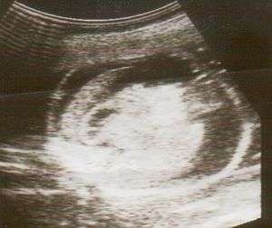 | |
| An ultrasound showing a fetus with Hydrops fetalis | |
| Specialty | Obstetrics and gynaecology, hematology, immunology |
Signs and symptoms
Locations can include:
- subcutaneous tissue/scalp
- pleura (pleural effusion)
- pericardium (pericardial effusion)
- abdomen (ascites)
The edema is usually seen in the fetal subcutaneous tissue, sometimes leading to spontaneous abortion. It is a prenatal form of heart failure, in which the heart is unable to satisfy demand (in most cases abnormally high) for blood flow.
Causes
Hydrops fetalis usually stems from fetal anemia, when the heart needs to pump a much greater volume of blood to deliver the same amount of oxygen. This anemia can have either an immune or non-immune cause. Non-immune hydrops can also be unrelated to anemia, for example if a fetal tumor or congenital cystic adenomatoid malformation increases the demand for blood flow. The increased demand for cardiac output leads to heart failure, and corresponding edema.
Immune
- Rh disease is an increasingly uncommon cause of immune-mediated hydrops fetalis. Due to preventative methods developed in the 1970s, the incidence of Rh disease has markedly declined. Rh disease can be prevented by administration of anti-D IgG (Rho(D) Immune Globulin) injections to RhD-negative mothers during pregnancy and/or within 72 hours of the delivery. However, a small percentage of pregnant mothers are still susceptible to Rh disease even after receiving anti-D IgG (Rho(D) Immune Globulin)
Non-immune
The non-immune form of hydrops fetalis has many causes including:[3]
- Iron deficiency anemia
- Paroxysmal supraventricular tachycardia resulting in heart failure
- Deficiency of the enzyme beta-glucuronidase. This enzyme deficiency is the cause of the lysosomal storage disease called mucopolysaccharidosis type VII.
- Congenital disorders of glycosylation
- Parvovirus B19 (fifth disease) infection of the pregnant woman
- Cytomegalovirus in mother
- Congenital pulmonary airway malformation (formerly called congenital cystic adenomatoid malformation)
- Maternal syphilis and maternal diabetes mellitus
- Alpha-thalassemia can also cause hydrops fetalis when all four of the genetic loci for α globin are deleted or affected by mutation. This is termed Hb Barts (consists of y-4 tetramers).
- Uncommonly, Niemann-Pick disease Type C (NPC) and Gaucher disease type 2 can present with hydrops fetalis.
- Turner syndrome
- Tumors,[4] the most common type of fetal tumor being teratoma, particularly a sacrococcygeal teratoma.
- Twin-twin transfusion syndrome in pregnancies in which twins share a single placenta (hydrops affects the recipient twin)
- Maternal hyperthyroidism
- Fetal cardiac defects and skeletal defects
- Noonan syndrome
- Mirror syndrome, in which fetal and placental hydrops develops in association with maternal preeclampsia, edema and hypertension
- Down syndrome
Diagnosis
Hydrops fetalis can be diagnosed and monitored by ultrasound scans. Prenatal ultrasound scanning enables early recognition of hydrops fetalis and has been enhanced with the introduction of MCA Doppler.
Treatment
The treatment depends on the cause.
Severely anemic fetuses, including those with Rh disease and alpha thalassemia major, can be treated with blood transfusions while still in the womb. This treatment increases the chance that the fetus will survive until birth.[3][5][6]
See also
References
- "Hydrops Fetalis: eMedicine Pediatrics: Cardiac Disease and Critical Care Medicine". Retrieved 2010-02-11.
- Knottenbelt, Derek C. (2003). Equine stud farm medicine and surgery. ISBN 9780702021305. Retrieved 2010-02-11.
- Norton, Mary E.; Chauhan, Suneet P.; Dashe, Jodi S. (2015-02-01). "Society for Maternal-Fetal Medicine (SMFM) Clinical Guideline #7: nonimmune hydrops fetalis". American Journal of Obstetrics and Gynecology. 212 (2): 127–139. doi:10.1016/j.ajog.2014.12.018. PMID 25557883.
- Isaacs H (January 2008). "Fetal hydrops associated with tumors". Am J Perinatol. 25 (1): 43–68. doi:10.1055/s-2007-1004826. PMID 18075961.
- Vichinsky, Elliott P. (2009-01-01). "Alpha thalassemia major—new mutations, intrauterine management, and outcomes". ASH Education Program Book. 2009 (1): 35–41. doi:10.1182/asheducation-2009.1.35. ISSN 1520-4391. PMID 20008180.
- Derderian, S. Christopher; Jeanty, Cerine; Fleck, Shannon R.; Cheng, Lily S.; Peyvandi, Shabnam; Moon-Grady, Anita J.; Farrell, Jody; Hirose, Shinjiro; Gonzalez, Juan (2015-01-01). "The many faces of hydrops". Journal of Pediatric Surgery. 50 (1): 50–54. doi:10.1016/j.jpedsurg.2014.10.027. PMC 4315667. PMID 25598092.
External links
| Classification | |
|---|---|
| External resources |