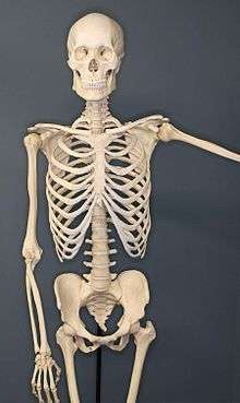
Human skeleton
The human skeleton is the internal framework of the human body. It is composed of around 270 bones at birth – this total decreases to around 206 bones by adulthood after some bones get fused together.[1] The bone mass in the skeleton reaches maximum density around age 21. The human skeleton can be divided into the axial skeleton and the appendicular skeleton. The axial skeleton is formed by the vertebral column, the rib cage, the skull and other associated bones. The appendicular skeleton, which is attached to the axial skeleton, is formed by the shoulder girdle, the pelvic girdle and the bones of the upper and lower limbs.
| Human Skeleton | |
|---|---|
| Details | |
| Identifiers | |
| Greek | σκελετός |
| TA | A02.0.00.000 |
| FMA | 23881 |
| Anatomical terminology | |
The human skeleton performs six major functions; support, movement, protection, production of blood cells, storage of minerals, and endocrine regulation.
The human skeleton is not as sexually dimorphic as that of many other primate species, but subtle differences between sexes in the morphology of the skull, dentition, long bones, and pelvis exist. In general, female skeletal elements tend to be smaller and less robust than corresponding male elements within a given population. The human female pelvis is also different from that of males in order to facilitate childbirth.[2] Unlike most primates, human males do not have penile bones.[3]
Skeletal divisions
Axial skeleton
The axial skeleton (80 bones) is formed by the vertebral column (32–34 bones; the number of the vertebrae differs from human to human as the lower 2 parts, sacral and coccygeal bone may vary in length), a part of the rib cage (12 pairs of ribs and the sternum), and the skull (22 bones and 7 associated bones).
The upright posture of humans is maintained by the axial skeleton, which transmits the weight from the head, the trunk, and the upper extremities down to the lower extremities at the hip joints. The bones of the spine are supported by many ligaments. The erector spinae muscles are also supporting and are useful for balance.
Appendicular skeleton
The appendicular skeleton (126 bones) is formed by the pectoral girdles, the upper limbs, the pelvic girdle or pelvis, and the lower limbs. Their functions are to make locomotion possible and to protect the major organs of digestion, excretion and reproduction.
Functions
The skeleton serves six major functions: support, movement, protection, production of blood cells, storage of minerals and endocrine regulation.
Support
The skeleton provides the framework which supports the body and maintains its shape. The pelvis, associated ligaments and muscles provide a floor for the pelvic structures. Without the rib cages, costal cartilages, and intercostal muscles, the lungs would collapse.
Movement
The joints between bones allow movement, some allowing a wider range of movement than others, e.g. the ball and socket joint allows a greater range of movement than the pivot joint at the neck. Movement is powered by skeletal muscles, which are attached to the skeleton at various sites on bones. Muscles, bones, and joints provide the principal mechanics for movement, all coordinated by the nervous system.
It is believed that the reduction of human bone density in prehistoric times reduced the agility and dexterity of human movement. Shifting from hunting to agriculture has caused human bone density to reduce significantly.[4][5][6]
Protection
The skeleton helps to protect our many vital internal organs from being damaged.
- The skull protects the brain
- The vertebrae protect the spinal cord.
- The rib cage, spine, and sternum protect the lungs, heart and major blood vessels.
Blood cell production
The skeleton is the site of haematopoiesis, the development of blood cells that takes place in the bone marrow. In children, haematopoiesis occurs primarily in the marrow of the long bones such as the femur and tibia. In adults, it occurs mainly in the pelvis, cranium, vertebrae, and sternum.[7]
Storage
The bone matrix can store calcium and is involved in calcium metabolism, and bone marrow can store iron in ferritin and is involved in iron metabolism. However, bones are not entirely made of calcium, but a mixture of chondroitin sulfate and hydroxyapatite, the latter making up 70% of a bone. Hydroxyapatite is in turn composed of 39.8% of calcium, 41.4% of oxygen, 18.5% of phosphorus, and 0.2% of hydrogen by mass. Chondroitin sulfate is a sugar made up primarily of oxygen and carbon.
Endocrine regulation
Bone cells release a hormone called osteocalcin, which contributes to the regulation of blood sugar (glucose) and fat deposition. Osteocalcin increases both the insulin secretion and sensitivity, in addition to boosting the number of insulin-producing cells and reducing stores of fat.[8]
Sex differences
Anatomical differences between human males and females are highly pronounced in some soft tissue areas, but tend to be limited in the skeleton. The human skeleton is not as sexually dimorphic as that of many other primate species, but subtle differences between sexes in the morphology of the skull, dentition, long bones, and pelvis are exhibited across human populations. In general, female skeletal elements tend to be smaller and less robust than corresponding male elements within a given population. It is not known whether or to what extent those differences are genetic or environmental.
Skull
A variety of gross morphological traits of the human skull demonstrate sexual dimorphism, such as the median nuchal line, mastoid processes, supraorbital margin, supraorbital ridge, and the chin.[9]
Dentition
Human inter-sex dental dimorphism centers on the canine teeth, but it is not nearly as pronounced as in the other great apes.
Long bones
Long bones are generally larger in males than in females within a given population. Muscle attachment sites on long bones are often more robust in males than in females, reflecting a difference in overall muscle mass and development between sexes. Sexual dimorphism in the long bones is commonly characterized by morphometric or gross morphological analyses.
Pelvis
The human pelvis exhibits greater sexual dimorphism than other bones, specifically in the size and shape of the pelvic cavity, ilia, greater sciatic notches, and the sub-pubic angle. The Phenice method is commonly used to determine the sex of an unidentified human skeleton by anthropologists with 96% to 100% accuracy in some populations.[10]
Women's pelvises are wider in the pelvic inlet and are wider throughout the pelvis to allow for child birth. The sacrum in the women's pelvis is curved inwards to allow the child to have a "funnel" to assist in the child's pathway from the uterus to the birth canal.
Clinical significance
There are many classified skeletal disorders. One of the most common is osteoporosis. Also common is scoliosis, a side-to-side curve in the back or spine, often creating a pronounced "C" or "S" shape when viewed on an x-ray of the spine. This condition is most apparent during adolescence, and is most common with females.
Arthritis
Arthritis is a disorder of the joints. It involves inflammation of one or more joints. When affected by arthritis, the joint or joints affected may be painful to move, may move in unusual directions or may be immobile completely. The symptoms of arthritis will vary differently between types of arthritis. The most common form of arthritis, osteoarthritis, can affect both the larger and smaller joints of the human skeleton. The cartilage in the affected joints will degrade, soften and wear away. This decreases the mobility of the joints and decreases the space between bones where cartilage should be.
Osteoporosis
Osteoporosis is a disease of bone where there is reduced bone mineral density, increasing the likelihood of fractures.[11] Osteoporosis is defined by the World Health Organization in women as a bone mineral density 2.5 standard deviations below peak bone mass, relative to the age and sex-matched average, as measured by Dual energy X-ray absorptiometry, with the term "established osteoporosis" including the presence of a fragility fracture.[12] Osteoporosis is most common in women after menopause, when it is called "postmenopausal osteoporosis", but may develop in men and premenopausal women in the presence of particular hormonal disorders and other chronic diseases or as a result of smoking and medications, specifically glucocorticoids.[11] Osteoporosis usually has no symptoms until a fracture occurs.[11] For this reason, DEXA scans are often done in people with one or more risk factors, who have developed osteoporosis and be at risk of fracture.[11]
Osteoporosis treatment includes advice to stop smoking, decrease alcohol consumption, exercise regularly, and have a healthy diet. Calcium supplements may also be advised, as may Vitamin D. When medication is used, it may include bisphosphonates, Strontium ranelate, and osteoporosis may be one factor considered when commencing Hormone replacement therapy.[11]
History
Sushruta, a famous medical scholar from India born in 600 BC, wrote the Suśruta-saṃhitā. In its extant form, its 184 chapters contain descriptions of 1,120 illnesses, 700 medicinal plants, 64 preparations from mineral sources and 57 preparations based on animal sources. The text discusses such surgical techniques as making incisions, probing, extraction of foreign bodies, alkali and thermal cauterization, tooth extraction, excisions, and trocars for draining abscess, draining hydrocele and ascitic fluid, removal of the prostate gland, urethral stricture dilatation, vesicolithotomy, hernia surgery, caesarian section, management of haemorrhoids, fistulae, laparotomy and management of intestinal obstruction, perforated intestines and accidental perforation of the abdomen with protrusion of omentum and the principles of fracture management, viz., traction, manipulation, apposition and stabilization including some measures of rehabilitation and fitting of prosthetic. It enumerates six types of dislocations, twelve varieties of fractures, and classification of the bones and their reaction to the injuries, and gives a classification of eye diseases including cataract surgery.
The study of bones in ancient Greece started under Ptolemaic kings due to their link to Egypt. Herophilos, through his work by studying dissected human corpses in Alexandria, is credited to be the pioneer of the field. His works are lost but are often cited by notable persons in the field such as Galen and Rufus of Ephesus. Galen himself did little dissection though and relied on the work of others like Marinus of Alexandria,[13] as well as his own observations of gladiator cadavers and animals.[14] According to Katherine Park, in medieval Europe dissection continued to be practiced, contrary to the popular understanding that such practices were taboo and thus completely banned.[15] The practice of holy autopsy, such as in the case of Clare of Montefalco further supports the claim.[16] Alexandria continued as a center of anatomy under Islamic rule, with Ibn Zuhr a notable figure. Chinese understandings are divergent, as the closest corresponding concept in the medicinal system seems to be the meridians, although given that Hua Tuo regularly performed surgery, there may be some distance between medical theory and actual understanding.
The Renaissance
Leonardo Da Vinci, among his many talents, also contributed to the study of the skeleton, albeit unpublished in his time.[17] Many artists, Antonio Pollaiuolo being the first, performed dissections for better understanding of the body, although they concentrated mostly on the muscles.[18] Vesalius, regarded as the founder of modern anatomy, authored the book De humani corporis fabrica, which contained many illustrations of the skeleton and other body parts, correcting some theories dating from Galen, such as the lower jaw being a single bone instead of two.[19] Various other figures like Alessandro Achillini also contributed to the further understanding of the skeleton.
References
| Library resources about Skeletal system |
- Mammal anatomy : an illustrated guide. New York: Marshall Cavendish. 2010. p. 129. ISBN 9780761478829.
- Thieme Atlas of Anatomy, (2006), p 113
- Patterns of Sexual Behavior Clellan S. Ford and Frank A. Beach, published by Harper & Row, New York in 1951. ISBN 0-313-22355-6
- "Switching Farming Made Human Bone Skeleton Joint Lighter". Smithsonian Magazine. 23 December 2014.
- "Light human skeleton may have come after agriculture". Retrieved 4 March 2017.
- "With the Advent of Agriculture, Human Bones Dramatically Weakened". 22 December 2014. Retrieved 4 March 2017.
- Fernández, KS; de Alarcón, PA (Dec 2013). "Development of the hematopoietic system and disorders of hematopoiesis that present during infancy and early childhood". Pediatric Clinics of North America. 60 (6): 1273–89. doi:10.1016/j.pcl.2013.08.002. PMID 24237971.
- Lee, Na Kyung; Sowa, Hideaki; Hinoi, Eiichi; Ferron, Mathieu; Ahn, Jong Deok; Confavreux, Cyrille; Dacquin, Romain; Mee, Patrick J.; McKee, Marc D.; Jung, Dae Young; Zhang, Zhiyou; Kim, Jason K.; Mauvais-Jarvis, Franck; Ducy, Patricia; Karsenty, Gerard (2007). "Endocrine Regulation of Energy Metabolism by the Skeleton". Cell. 130 (3): 456–69. doi:10.1016/j.cell.2007.05.047. PMC 2013746. PMID 17693256.
- Buikstra, J.E.; D.H. Ubelaker (1994). Standards for data collection from human skeletal remains. Arkansas Archaeological Survey. p. 208.
- Phenice, T. W. (1969). "A newly developed visual method of sexing the os pubis". American Journal of Physical Anthropology. 30 (2): 297–301. doi:10.1002/ajpa.1330300214. PMID 5772048.
- Britton, the editors Nicki R. Colledge, Brian R. Walker, Stuart H. Ralston; illustrated by Robert (2010). Davidson's principles and practice of medicine (21st ed.). Edinburgh: Churchill Livingstone/Elsevier. pp. 1116–1121. ISBN 978-0-7020-3085-7.
- WHO (1994). "Assessment of fracture risk and its application to screening for postmenopausal osteoporosis. Report of a WHO Study Group". World Health Organization technical report series. 843: 1–129. PMID 7941614.
- Rocca, Julius (9 August 2010). "A Note on Marinus of Alexandria". Journal of the History of the Neurosciences. 11 (3): 282–285. doi:10.1076/jhin.11.3.282.10386.
- Charlier, Philippe; Huynh-Charlier, Isabelle; Poupon, Joël; Lancelot, Eloïse; Campos, Paula F.; Favier, Dominique; Jeannel, Gaël-François; Bonati, Maurizio Rippa; Grandmaison, Geoffroy Lorin de la; Herve, Christian (2014). "Special report: Anatomical pathology A glimpse into the early origins of medieval anatomy through the oldest conserved human dissection (Western Europe, 13th c. A.D.)". Archives of Medical Science. 2: 366–373. doi:10.5114/aoms.2013.33331. PMC 4042035.
- "Debunking a myth". Harvard Gazette. Retrieved 12 November 2016.
- Hairston, Julia L.; Stephens, Walter (2010). The body in early modern Italy. Baltimore: Johns Hopkins University Press. ISBN 9780801894145.
- Sooke, Alastair. "Leonardo da Vinci: Anatomy of an artist". Telegraph.co.uk. Retrieved 9 December 2016.
- Bambach, Carmen. "Anatomy in the Renaissance". The Met’s Heilbrunn Timeline of Art History.
- "Vesalius's Renaissance anatomy lessons". www.bl.uk. Retrieved 18 December 2016.
| Authority control |
|
|---|