Hindbrain
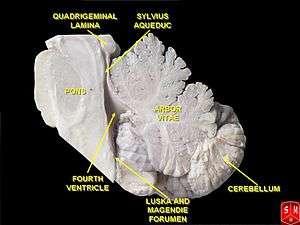
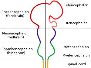
The hindbrain or rhombencephalon is a developmental categorization of portions of the central nervous system in vertebrates. It includes the medulla, pons, and cerebellum. Together they support vital bodily processes.[1]
| Hindbrain | |
|---|---|
 | |
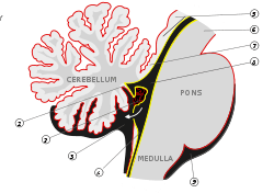 Scheme of roof of fourth ventricle. | |
| Identifiers | |
| MeSH | D012249 |
| NeuroNames | 540 |
| NeuroLex ID | birnlex_942 |
| TA | A14.1.03.002 |
| FMA | 67687 |
| Anatomical terms of neuroanatomy | |
The hindbrain can be subdivided in a variable number of transversal swellings called rhombomeres. In the human embryo eight rhombomeres can be distinguished, from caudal to rostral: Rh8-Rh1. Rostrally, the isthmus demarcates the boundary with the midbrain.
The caudal rhombencephalon has been generally considered as the initiation site for neural tube closure.[2]
Metencephalon
Rhombomeres Rh3-Rh1 form the metencephalon.
The metencephalon is composed of the pons and the cerebellum; it contains:
- a portion of the fourth(IV) ventricle,
- the trigeminal nerve (CN V),
- abducens nerve (CN VI),
- facial nerve (CN VII),
- and a portion of the vestibulocochlear nerve (CN VIII).
Myelencephalon
Rhombomeres Rh8-Rh4 form the myelencephalon.
The myelencephalon forms the medulla oblongata in the adult brain; it contains:
- a portion of the fourth ventricle,
- the glossopharyngeal nerve (CN IX),
- vagus nerve (CN X),
- accessory nerve (CN XI),
- hypoglossal nerve (CN XII),
- and a portion of the vestibulocochlear nerve (CN VIII).
Evolution
The hindbrain is homologous to a part of the arthropod brain known as the sub-oesophageal ganglion, in terms of the genes that it expresses and its position in between the brain and the nerve cord.[3] On this basis, it has been suggested that the hindbrain first evolved in the Urbilaterian—the last common ancestor of chordates and arthropods—between 570 and 555 million years ago.[3][4]
Hindbrain diseases
A rare brain malformation of the cerebellum is rhombencephalosynapsis characterized by an absent or partially formed vermis. Symptoms can include ataxia. The disorder is a main feature of Gomez-Lopez-Hernandez syndrome.
Additional images
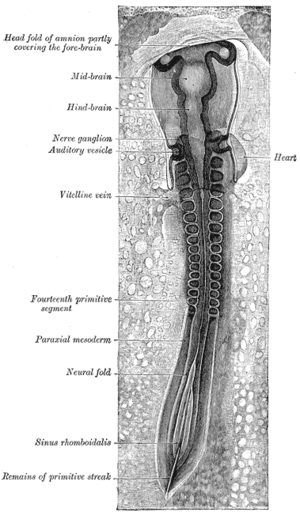 Chicken embryo of thirty-three hours’ incubation, viewed from the dorsal aspect. X 30.
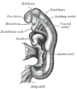 Human embryo between eighteen and twenty-one days.
 Hindbrain of human embryo
References
- Haycock DE (2011). Being and Perceiving. Manupod Press. p. 41. ISBN 978-0-9569621-0-2.
- "Brain atlas - Hindbrain". Lundbeck Institute - Brain explorer. Retrieved 2015-06-08.
- SpringerLink - Journal Article
- Ghysen A (2003). "The origin and evolution of the nervous system". Int. J. Dev. Biol. 47 (7–8): 555–62. PMID 14756331.
- Haycock, DE Being and Perceiving
5. Gisele E. Ishak, Jennifer C. Dempsey, Dennis W. W. Shaw, Hannah Tully, Margaret P. Adam, Pedro A. Sanchez-Lara, Ian Glass, Tessa C. Rue, Kathleen J. Millen, William B. Dobyns, Dan Doherty; Rhombencephalosynapsis: a hindbrain malformation associated with incomplete separation of midbrain and forebrain, hydrocephalus, and a broad spectrum of severity, Brain, Volume 135, Issue 5, 1 May 2012, Pages 1370-1386, https://doi.org/10.1093/brain/aws065
6. Tully, H. M., Dempsey, J. C., Ishak, G. E., Adam, M. P., Mink, J. W., Dobyns, W. B., Gospe, S. M., Weiss, A., Phillips, J. O. and Doherty, D. (2013), Persistent figure‐eight and side‐to‐side head shaking is a marker for rhombencephalosynapsis. Mov Disord., 28: 2019-2023. doi:10.1002/mds.25634
7. Poretti, Andrea & Dietrich Alber, Fabienne & Buerki, Sarah & P Toelle, Sandra & Boltshauser, Eugen. (2008). Cognitive outcome in children with rhombencephalosynapsis. European journal of paediatric neurology : EJPN : official journal of the European Paediatric Neurology Society. 13. 28-33. 10.1016/j.ejpn.2008.02.005.
8. D Bell, Brian & A Stanko, Heather & L Levine, Ross. (2005). Normal IQ in a 55-year-old with newly diagnosed rhombencephalosynapsis. Archives of clinical neuropsychology : the official journal of the National Academy of Neuropsychologists. 20. 613-21. 10.1016/j.acn.2005.02.003.
External links
| Look up hindbrain in Wiktionary, the free dictionary. |
- NIF Search - Hindbrain via the Neuroscience Information Framework
| Authority control |
|
|---|