Granulosa cell tumour
Granulosa cell tumours are tumours that arise from granulosa cells. They are esterogen secreting tumors and present as large, complex, ovarian masses. These tumours are part of the sex cord-gonadal stromal tumour or non-epithelial group of tumours. Although granulosa cells normally occur only in the ovary, granulosa cell tumours occur in both ovaries and testicles (see Ovarian cancer and Testicular cancer). These tumours should be considered malignant and treated in the same way as other malignant tumours of ovary. The ovarian disease has two forms, juvenile and adult, both characterized by indolent growth,[1] and therefore has high recovery rates.[2][3] The staging system for these tumours is the same as for epithelial tumours and most present as stage I.[4] The peak age at which they occur is 50–55 years, but they may occur at any age.
| Granulosa cell tumour | |
|---|---|
| Other names | Granulosa-theca cell tumours or Folliculoma |
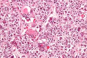 | |
| Micrograph of a juvenile granulosa cell tumour with hyaline globules. H&E stain. | |
| Specialty | Gynecologic oncology, obstetrics and gynaecology, oncology, endocrinology |
Juvenile granulosa cell tumour is a similar but distinct rare tumour. It too occurs in both the ovary and testis. In the testis it is extremely rare, and has not been reported to be malignant.[5] Although this tumour usually occurs in children (hence its name), it has been reported in adults.[6]
Presentation
Estrogens are produced by functioning tumours, and the clinical presentation depends on the patient's age and sex.
- Female
- If the patient is postmenopausal, she usually presents with abnormal uterine bleeding, and in some cases hemoperitoneum.
- If the patient is of reproductive age, she would present with menometrorrhagia. However, in some cases she may stop ovulating altogether.
- If the patient has not undergone puberty, early onset of puberty may be seen.
- these tumors tend to have late recurrencies ( even after 30 years )
Genetics
Adult Granulosa Cell Tumors
Using next generation DNA sequencing, it was discovered that 97% of adult granulosa cell tumours contain an identical mutation in the FOXL2 gene . This is a somatic mutation, meaning it is not usually transmitted to descendants. Mutation c.402C>G in the sequence of FOXL2 leads to the amino acid substitution p. C134W. It is believed that this mutation may be the cause of granulosa cell tumours.
Juvenile Granulosa Cell Tumors
Two recent studies show that the enzyme AKT1 is involved in juvenile granulosa cell tumors. In-frame duplications in the pleckstrin-homology domain of the protein were found in more than 60% of juvenile granulosa cell tumors occurring in girls under 15 years of age. The tumors without duplications carried point mutations affecting highly conserved residues. The mutated proteins carrying the duplications displayed a non-wild-type subcellular distribution, with a marked enrichment at the plasma membrane. This led to a striking degree of AKT1 activation demonstrated by a strong phosphorylation level and corroborated by reporter assays.[7]
Analysis by RNA-Seq pinpointed a series of differentially expressed genes, involved in cytokine and hormone signaling and cell division-related processes. Further analyses pointed to a possible dedifferentiation process and suggested that most of the transcriptomic dysregulations might be mediated by a limited set of transcription factors perturbed by AKT1 activation. These results incriminate somatic mutations of AKT1 as major probably driver events in the pathogenesis of juvenile granulosa cell tumors.[8]
Diagnosis
Gross appearance
Tumors vary in size, from tiny spots to large masses, with an average of 10 cm in diameter. Tumors are oval and soft in consistency. On cut-section, histology reveals reticular, trabecular areas with interstitial haemorrhage and Call-Exner bodies-small cyst like spaces interspersed within a graafian follicle.
Tumor marker
Inhibin, a hormone, has been used as tumor marker for granulosa cell tumor.
Treatment
In animals
In the ovaries of aging squirrel monkeys (Saimiri sciureus), clusters of granulosa cells occur that resemble granulosa cell tumours in humans.[9] These appear to be a normal change with age in this species.
See also
- Inhibin, alpha
References
- "Prognostic factors in adult granulosa".
- Young RH, Dickersin GR, Scully RE (1984). "Juvenile granulosa cell tumor of the ovary. A clinic pathological analysis of 125 cases. Beth Israel Deaconess Medical Center, Boston". American Journal of Surgical Pathology. 8 (8): 575–596. doi:10.1097/00000478-198408000-00002. PMID 6465418.
- "Program in Gynecologic Medical Oncology, Beth Israel Deaconess Medical Center, Boston".
- Gynaecology. 3rd Ed. 2003. Churchill Livingstone, pp. 690-691.
- Dudani R, Giordano L, Sultania P, Jha K, Florens A, Joseph T (April 2008). "Juvenile granulosa cell tumor of testis: case report and review of literature". American Journal of Perinatology. 25 (4): 229–231. doi:10.1055/s-2008-1066878. PMID 18548396.
- Lin KH, Lin SE, Lee LM (July 2008). "Juvenile granulosa cell tumor of adult testis: a case report". Urology. 72 (1): 230.e11–3. doi:10.1016/j.urology.2007.11.126. PMID 18313118.
- Bessière L, Todeschini AL, Auguste A, Sarnacki S, Flatters D, Legois B, Sultan C, Kalfa N, Galmiche L, Veitia RA (March 2015). "A Hot-spot of In-frame Duplications Activates the Oncoprotein AKT1 in Juvenile Granulosa Cell Tumors". EBioMedicine. 2 (5): 421–431. doi:10.1016/j.ebiom.2015.03.002. PMC 4485906. PMID 26137586.
- Auguste A, Bessière L, Todeschini AL, Caburet S, Sarnacki S, Prat J, D'angelo E, De La Grange P, Ariste O, Lemoine F, Legois B, Sultan C, Zider A, Galmiche L, Kalfa N, Veitia RA (Dec 2015). "Molecular analyses of juvenile granulosa cell tumors bearing AKT1 mutations provide insights into tumor biology and therapeutic leads". Human Molecular Genetics. 24 (23): 6687–6698. doi:10.1093/hmg/ddv373. PMID 26362254.
- Walker ML, Anderson DC, Herndon JG, Walker LC (2009). "Ovarian aging in squirrel monkeys (Saimiri sciureus)". Reproduction. 138 (4): 793–799. doi:10.1530/REP-08-0449. PMID 19656956.
External links
| Classification | |
|---|---|
| External resources |