Granular cell tumor
Granular cell tumor is a tumor that can develop on any skin or mucosal surface, but occurs on the tongue 40% of the time.
| Granular cell tumor | |
|---|---|
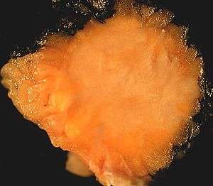 | |
| 2-cm tumor presented as an abdominal wall mass in a middle-aged woman | |
| Specialty | Oncology |
_skin.jpg)
It is also known as Abrikossoff's tumor,[1] Granular cell myoblastoma,[1] Granular cell nerve sheath tumor,[1] and Granular cell schwannoma.[1])Granular cell tumors (GCTs) affect females more often than males.[2]
Pathology
Granular cell tumors show similarity to neural tissue, as can be demonstrated by immunohistochemistry and ultrastructural evidence using electron microscopy.
Multiple granular cell tumors may seen in the context of LEOPARD syndrome, due to a mutation in the PTPN11 gene.[3]
These tumors on occasion may appear similar to neoplasms of renal (relating to the kidneys) origin or other soft tissue neoplasms.
Treatment
The primary method for treatment is surgical, not medical. Radiation and chemotherapy are not needed for benign lesions and are not effective for malignant lesions.
Benign granular cell tumors have a recurrence rate of 2% to 8% when resection margins are deemed clear of tumor infiltration. When the resection margins of a benign granular cell tumor are positive for tumor infiltration the recurrence rate is increased to 20%. Malignant lesions are aggressive and difficult to eradicate with surgery and have a recurrence rate of 32%.
Epidemiology
Granular cell tumors can affect all parts of the body; however, the head and neck areas are affected 45% to 65% of the time. Of the head and neck cases 70% of lesions are located intraorally (tongue, oral mucosa, hard palate). The next most common location that lesions are found in the head and neck area is the larynx (10%).[4] Granular cell tumors are also found in the internal organs, particularly in the upper aerodigestive tract. Vaginal granular cell tumor is generally rare.[5]
The usual presentation is of slow growing behavior, forming a polygonal accumulation of secondary lysosomes in the cytoplasm. Granular cell tumors are typically solitary and are rarely larger than three centimeters. This type of tumor has been found to be both benign and malignant, although malignancy is rare and comprises only 2% of all granular cell tumors.[6]
References
- Rapini, Ronald P.; Bolognia, Jean L.; Jorizzo, Joseph L. (2007). Dermatology: 2-Volume Set. St. Louis: Mosby. pp. 1796, 1804. ISBN 978-1-4160-2999-1.
- "Granular cell tumor | Genetic and Rare Diseases Information Center (GARD) – an NCATS Program". Genetic and rare diseases information center. Retrieved 17 April 2018.
- Schrader, KA.; Nelson, TN.; De Luca, A.; Huntsman, DG.; McGillivray, BC. (Feb 2009). "Multiple granular cell tumors are an associated feature of LEOPARD syndrome caused by mutation in PTPN11". Clin Genet. 75 (2): 185–9. doi:10.1111/j.1399-0004.2008.01100.x. PMID 19054014.
- Kahn, Michael A. Basic Oral and Maxillofacial Pathology. Volume 1. 2001.
- Danforth's obstetrics and gynecology. Gibbs, Ronald S., 1943-, Danforth, David N. (David Newton), 1912-1990. (10th ed.). Philadelphia: Lippincott Williams & Wilkins. 2008. ISBN 9780781769372. OCLC 187289377.CS1 maint: others (link)
- Fanburg-Smith JC, Meis-Kindblom JM, et al. Malignant granular cell tumor of soft tissue: diagnostic criteria and clinicopathologic correlation. Am J Surg Pathol. Jul 1998;22(7):779-94.
External links
| Classification |
|---|