Golgi cell
In neuroscience, Golgi cells are inhibitory interneurons found within the granular layer of the cerebellum. They were first identified as inhibitory by Eccles et al. in 1964.[1] It was also the first example of an inhibitory feed back network, where the inhibitory interneuron was identified anatomically. These cells synapse onto the dendrite of granule cells and unipolar brush cells. They receive excitatory input from mossy fibres, also synapsing on granule cells, and parallel fibers, which are long granule cell axons. Thereby this circuitry allows for feed-forward and feed-back inhibition of granule cells.
| Golgi cell | |
|---|---|
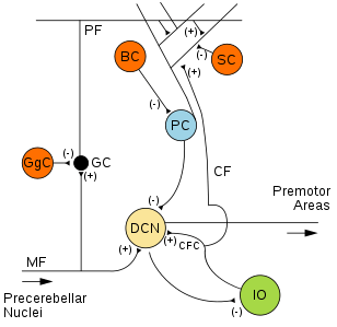 Microcircuitry of the cerebellum. Excitatory synapses are denoted by (+) and inhibitory synapses by (-). MF: Mossy fiber. DCN: Deep cerebellar nuclei. IO: Inferior olive. CF: Climbing fiber. GC: Granule cell. PF: Parallel fiber. PC: Purkinje cell. GgC: Golgi cell. SC: Stellate cell. BC: Basket cell. | |
| Details | |
| Location | Granular layer of the cerebellum |
| Identifiers | |
| NeuroLex ID | nifext_129 |
| Anatomical terms of neuroanatomy | |
The main synapse made by these cells is a synapse onto the mossy fibre - granule cell excitatory synapse in a glomerulus. The glomerulus is made up of the mossy fibre terminal, granule cell dendrites, the Golgi terminal and is enclosed by a glial coat.[2] The Golgi cell acts by altering the mossy fibre - granule cell synapse.
The Golgi cells use GABA as their transmitter. The basal level of GABA produces a postsynaptic leak conductance by tonically activating alpha 6-containing GABA-A receptors on the granule cell.[3][4][5] These high-affinity receptors are located both synaptically and extrasynaptically on the granule cell. The synaptic receptors mediate phasic contraction, duration of around 20-30ms whereas the extrasynapatic receptors mediate tonic inhibition of around 200ms, and are activated by synapse spill over.[6]
Additionally the GABA acts on GABA-B receptors which are located presynaptically on the mossy fibre terminal. These inhibit the mossy fibre evoked EPSCs of the granule cell in a temperature and frequency dependent manner. At high mossy firing frequency (10 Hz) there is no effect of GABA acting on presynaptic GABA-B receptors on evoked EPSCs. However, at low (1 Hz) firing the GABA does have an effect on the EPSCs mediated via these presynaptic GABA-B receptors.
References
- Eccles, JC; Llinas, R; Sasaki, K (1964). "Golgi cell inhibition in the cerebellar cortex". Nature. 204 (4965): 1265–1266. doi:10.1038/2041265a0. PMID 14254404.
- Jakab, RL; Hámori, J (1988). "Quantitative morphology and synaptology of cerebellar glomeruli in the rat". Anatomy and Embryology. 179 (100): 81–88. doi:10.1007/BF00305102. PMID 3213958.
- Brickley SG, Cull-Candy SG, Farrant M (1996). "Development of a tonic form of synaptic inhibition in rat cerebellar granule cells resulting from persistent activation of GABAA receptors". J Physiol. 497 (Pt 3): 753–759. doi:10.1113/jphysiol.1996.sp021806. PMC 1160971. PMID 9003560.
- Tia S, Wang JF, Kotchabhakdi N, Vicini S (June 1, 1996). "Developmental changes of inhibitory synaptic currents in cerebellar granule neurons: role of GABAA receptor alpha 6 subunit" (abstract). Journal of Neuroscience. 16 (11): 3630–3640. doi:10.1523/JNEUROSCI.16-11-03630.1996. PMID 8642407.
- Wall MJ, Usowicz MM (1997). "Development of action potential-dependent and independent spontaneous GABAA receptor-mediated currents in granule cells of postnatal rat cerebellum". European Journal of Neuroscience. 9 (3): 533–548. doi:10.1111/j.1460-9568.1997.tb01630.x. PMID 9104595.
- Nusser Z, Sieghart W, Somogyi P (March 1, 1998). "Segregation of different GABAA receptors to synaptic and extrasynaptic membranes of cerebellar granule cells" (abstract). Journal of Neuroscience. 18 (5): 1693–1703. PMID 9464994.
External links
- NIF Search - Golgi Cell via the Neuroscience Information Framework