Francisella tularensis
Francisella tularensis is a pathogenic species of Gram-negative coccobacillus, an aerobic bacterium.[1] It is nonspore-forming, nonmotile,[2] and the causative agent of tularemia, the pneumonic form of which is often lethal without treatment. It is a fastidious, facultative intracellular bacterium, which requires cysteine for growth.[3] Due to its low infectious dose, ease of spread by aerosol, and high virulence, F. tularensis is classified as a Tier 1 Select Agent by the U.S. government, along with other potential agents of bioterrorism such as Yersinia pestis, Bacillus anthracis, and Ebola virus. When found in nature, Francisella tularensis can survive for several weeks at low temperatures in animal carcasses, soil, and water. In the laboratory, F. tularensis appears as small rods (0.2 by 0.2 µm), and is grown best at 35-37°C.[4]
| Francisella tularensis | |
|---|---|
.jpg) | |
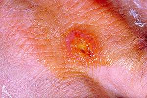
| Francisella tularensis bacteria (blue) infecting a macrophage (yellow) | |
| Scientific classification | |
| Domain: | Bacteria |
| Phylum: | |
| Class: | |
| Order: | |
| Family: | Francisellaceae |
| Genus: | |
| Species: | F. tularensis |
| Binomial name | |
| Francisella tularensis (McCoy and Chapin 1912) Dorofe'ev 1947 | |
Subspecies
This species was discovered in ground squirrels in Tulare County, California, in 1911; Bacterium tularense was soon isolated by George Walter McCoy (1876–1952) of the US Plague Lab in San Francisco and reported in 1912. In 1922, Dr. Edward Francis (1872–1957), a physician and medical researcher from Ohio, discovered that Bacterium tularense was the causative agent of tularemia, after studying several cases of his patients having symptoms of the said disease. Later, it became known as "Francisella tularensis", in honor of the discovery by Dr. Francis.[5][6][7] Four subspecies (biovars) of F. tularensis have been classified:
- F. t. tularensis (or type A), found predominantly in North America, is the most virulent of the four known subspecies, and is associated with lethal pulmonary infections. This includes the primary type A laboratory strain, SCHUS4.
- F. t. holarctica (also known as biovar F. t. palearctica or type B) is found predominantly in Europe and Asia, but rarely leads to fatal disease. An attenuated live vaccine strain of subspecies F. t. holarctica has been described, though it is not yet fully licensed by the Food and Drug Administration as a vaccine. This subspecies lacks the citrulline ureidase activity and ability to produce acid from glucose of biovar F. t. palearctica.
- F. t. novicida (previously classified as F. novicida[8]) was characterized as a relatively nonvirulent strain; only two tularemia cases in North America have been attributed to F. t. novicida and these were only in severely immunocompromised individuals.
- F. t. mediasiatica, is found primarily in central Asia; little is currently known about this subspecies or its ability to infect humans.
In 1938, Soviet bacteriologist Vladimir Dorofeev (1911–1988) and his team were able to recreate the infectious cycle of the pathogen in humans, and his team was the first in the world to create measures in protection against the deadly infectious agent. In 1947, Dorofeev was able to independently isolate the pathogen that Dr. Francis discovered in 1922; hence, it is commonly known as Francisella dorofeev in former Soviet countries.
Pathogenesis
F. tularensis has been reported in birds, reptiles, fish, invertebrates, and mammals including humans. Despite this, no case of tularemia has been shown to be initiated by human-to-human transmission. Rather, it is caused by contact with infected animals or vectors such as ticks, mosquitos, and deer flies. Reservoir hosts of importance can include lagomorphs (e.g. rabbits), rodents, galliform birds, and deer.
Infection with F. tularensis can occur by several routes. Portals of entry are through blood and the respiratory system. The most common occurs via skin contact, yielding an ulceroglandular form of the disease. Inhalation of bacteria - particularly biovar F. t. tularensis, leads to the potentially lethal pneumonic tularemia. While the pulmonary and ulceroglandular forms of tularemia are more common, other routes of inoculation have been described and include oropharyngeal infection due to consumption of contaminated food and conjunctival infection due to inoculation at the eye.
F. tularensis is capable of surviving outside of a mammalian host for weeks at a time and has been found in water, grassland, and haystacks. Aerosols containing the bacteria may be generated by disturbing carcasses due to brush cutting or lawn mowing; as a result, tularemia has been referred to as "lawnmower disease". Recent epidemiological studies have shown a positive correlation between occupations involving the above activities and infection with F. tularensis.
Lifecycle
F. tularensis is a facultative intracellular bacterium that is capable of infecting most cell types, but primarily infects macrophages in the host organism. Entry into the macrophage occurs by phagocytosis and the bacterium is sequestered from the interior of the infected cell by a phagosome. F. tularensis then breaks out of this phagosome into the cytosol and rapidly proliferates. Eventually, the infected cell undergoes apoptosis, and the progeny bacteria are released to initiate new rounds of infection.
Virulence factors
The virulence mechanisms for F. tularensis have not been well characterized. Like other intracellular bacteria that break out of phagosomal compartments to replicate in the cytosol, F. tularensis strains produce different hemolytic agents, which may facilitate degradation of the phagosome.[9] A hemolysin activity, named NlyA, with immunological reactivity to Escherichia coli anti-HlyA antibody, was identified in biovar F. t. novicida.[10] Acid phosphatase AcpA has been found in other bacteria to act as a hemolysin, whereas in Francisella, its role as a virulence factor is under vigorous debate.
F. tularensis contains type VI secretion system (T6SS), also present in some other pathogenic bacteria.[11] It also contains a number of ATP-binding cassette (ABC) proteins that may be linked to the secretion of virulence factors.[12] F. tularensis uses type IV pili to bind to the exterior of a host cell and thus become phagocytosed. Mutant strains lacking pili show severely attenuated pathogenicity.
The expression of a 23-kD protein known as IglC is required for F. tularensis phagosomal breakout and intracellular replication; in its absence, mutant F. tularensis cells die and are degraded by the macrophage. This protein is located in a putative pathogenicity island regulated by the transcription factor MglA.
F. tularensis, in vitro, downregulates the immune response of infected cells, a tactic used by a significant number of pathogenic organisms to ensure their replication is (albeit briefly) unhindered by the host immune system by blocking the warning signals from the infected cells. This downmodulation of the immune response requires the IglC protein, though again the contributions of IglC and other genes are unclear. Several other putative virulence genes exist, but have yet to be characterized for function in F. tularensis pathogenicity.
Genetics
Like many other bacteria, F. tularensis undergoes asexual replication. Bacteria divide into two daughter cells, each of which contains identical genetic information. Genetic variation may be introduced by mutation or horizontal gene transfer.
The genome of F. t. tularensis strain SCHU4 has been sequenced.[13] The studies resulting from the sequencing suggest a number of gene-coding regions in the F. tularensis genome are disrupted by mutations, thus create blocks in a number of metabolic and synthetic pathways required for survival. This indicates F. tularensis has evolved to depend on the host organism for certain nutrients and other processes ordinarily taken care of by these disrupted genes.
The F. tularensis genome contains unusual transposon-like elements resembling counterparts that normally are found in eukaryotic organisms.
Phylogenetics
Much of the known global genetic diversity of F. t. holarctica is present in Sweden.[14] This suggests this subspecies originated in Scandinavia and spread from there to the rest of Eurosiberia.
Use as a biological weapon
When the U.S. biological warfare program ended in 1969, F. tularensis was one of seven standardized biological weapons it had developed.[15]
Diagnosis, treatment, and prevention

- Diagnosis
Infection by F. tularensis is diagnosed by clinicians based on symptoms and patient history, imaging, and laboratory studies.
- Treatment
Tularemia is treated with antibiotics, such as aminoglycosides, tetracyclines, or fluoroquinolones. About 15 proteins were suggested that could facilitate drug and vaccine design pipeline.[16]
- Prevention
Preventive measures include preventing bites from ticks, flies, and mosquitos; ensuring that all game is cooked thoroughly; refraining from drinking untreated water; using insect repellents; if working with cultures of F. tularensis, in the lab, making sure to wear a gown, impermeable gloves, mask, and eye protection; and when dressing game, making sure to wear impermeable gloves. Also, a live attenuated vaccine is available for individuals who are at high risk for exposure such as laboratory personnel.[17]
Genomics
See also
- Francisella small RNA
References
- "Francisella tularensis" (PDF). health.ny.gov. Wadsworth Center: New York State Department of Health. Retrieved 12 May 2015.
- "Tularemia (Francisella tularensis)" (PDF). michigan.gov. Michigan Department of Community Health. Retrieved 12 May 2015.
- Ryan KJ; Ray CG (editors) (2004). Sherris Medical Microbiology (4th ed.). McGraw Hill. pp. 488–90. ISBN 0-8385-8529-9.CS1 maint: extra text: authors list (link)
- "Francisella tularensis - microbewiki".
- A. Tärnvik1 and L. Berglund, Tularaemia. Eur Respir J 2003; 21:361-373.
- McCoy GW, Chapin CW. Bacterium tularense, the cause of a plaguelike disease of rodents. Public Health Bull 1912;53:17–23.
- Jeanette Barry, Notable Contributions to Medical Research by Public Health Service Scientists. National Institute of Health, Public Health Service Publication No. 752, 1960, p. 36.
- Sjöstedt AB. "Genus I. Francisella Dorofe'ev 1947, 176AL". Bergey's Manual of Systematic Bacteriology. 2 (The Proteobacteria), part B (The Gammaproteobacteria) (2nd ed.). New York: Springer. pp. 200–210.
- http://iai.asm.org/cgi/reprint/76/8/3690
- Wiley Interscience
- Spidlova, Petra; Stulik, Jiri (2017). "Francisella tularensis type VI secretion system comes of age". Virulence. 8 (6): 628–631. doi:10.1080/21505594.2016.1278336. ISSN 2150-5594. PMC 5626347. PMID 28060573.
- Atkins H, Dassa E, Walker N, Griffin K, Harland D, Taylor R, Duffield M, Titball R (2006). "The identification and evaluation of ATP binding cassette systems in the intracellular bacterium Francisella tularensis". Res Microbiol. 157 (6): 593–604. doi:10.1016/j.resmic.2005.12.004. PMID 16503121.
- Larsson P, Oyston P, Chain P, et al. (2005). "The complete genome sequence of Francisella tularensis, the causative agent of tularemia". Nat Genet. 37 (2): 153–9. doi:10.1038/ng1499. PMID 15640799.
- Karlsson E, Svensson K, Lindgren P, Byström M, Sjödin A, Forsman M, Johansson A (2012) The phylogeographic pattern of Francisella tularensis in Sweden indicates a Scandinavian origin of Eurosiberian tularaemia. Environ Microbiol doi: 10.1111/1462-2920.12052
- Croddy, Eric C. and Hart, C. Perez-Armendariz J., Chemical and Biological Warfare, (Google Books), Springer, 2002, pp. 30-31, (ISBN 0387950761), accessed October 24, 2008.
- Francisella tularensis: In silico Identification of Drug and Vaccine Targets by Metabolic Pathway Analysis J Harati, J Fallah The 6th Conference on Bioinformatics
- http://publichealth.lacounty.gov/acd/procs/b73/B73Part4.pdf
External links
- Francisella tularensis information from the CDC/National Center for Infectious Diesase:
- BioHealthBase Bioinformatics Resource Center The National Institute of Allergy and Infectious Disease (NIAID) supports a public database describing the molecular genetics of F. tularensis. The website describes the genes, proteins, and cellular characteristics of the pathogen.
- Type strain of Francisella tularensis at BacDive - the Bacterial Diversity Metadatabase