Joint capsule
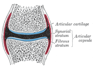
In anatomy, a joint capsule or articular capsule is an envelope surrounding a synovial joint.[1] Each joint capsule has two parts: an outer fibrous layer or membrane, and an inner synovial layer or membrane.
| Joint capsule | |
|---|---|
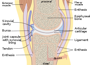 Typical joint | |
 Diagrammatic section of a diarthrodial joint. | |
| Details | |
| Identifiers | |
| Latin | capsula articuno |
| MeSH | D017746 |
| TA | A03.0.00.026 |
| FMA | 34836 |
| Anatomical terminology | |
Membranes
Each capsule consists of two layers or meme:
- an outer (fibrous membrane, fibrous stratum) composed of avascular white fibrous tissue
- an inner (synovial membrane, synovial stratum) which is a secreting layer
On the inside of the capsule, articular cartilage covers the end surfaces of the bones that articulate within that joint.
The outer layer is highly innervated by the same nerves which perforate through the adjacent muscles associated with the joint.
Fibrous membrane
The fibrous membrane of the joint capsule is attached to the whole circumference of the articular end of each bone entering into the joint, and thus entirely surrounds the articulation. It is made up of dense irregular connective tissue. It's a long spongy tissue.
Clinical significance
Frozen shoulder (adhesive capsulitis) is a disorder in which the shoulder capsule becomes inflamed.
Plica syndrome is a disorder in which the synovial plica becomes inflamed and causes abnormal biomechanics in the knee.
Gallery
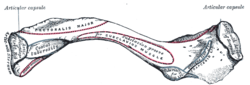 Left clavicle. Inferior surface.
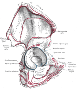 Right hip bone. External surface.
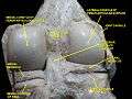 Right knee in extension. Deep dissection. Posterior view.
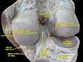 Right knee in extension. Deep dissection. Posterior view.
See also
References
This article incorporates text in the public domain from page 282 of the 20th edition of Gray's Anatomy (1918)
- eMedicine/Stedman Medical Dictionary Lookup! Archived 2008-04-12 at the Wayback Machine
External links
- Cross section image: pelvis/pelvis-e12-15—Plastination Laboratory at the Medical University of Vienna
| Authority control |
|---|