Fibrocartilage
White fibrocartilage consists of a mixture of white fibrous tissue and cartilaginous tissue in various proportions. It owes its inflexibility and toughness to the former of these constituents, and its elasticity to the latter. It is the only type of cartilage that contains Type I collagen in addition to the normal type II.
| Fibrocartilage | |
|---|---|
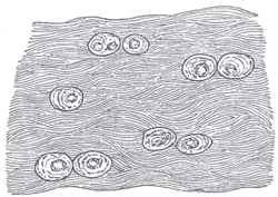 White fibrocartilage from an intervertebral fibrocartilage. | |
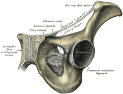 Symphysis pubis exposed by a coronal section. (Pubic symphysis visible at center left.) | |
| Identifiers | |
| MeSH | D051445 |
| TH | H2.00.03.5.00017 |
| Anatomical terminology | |
Fibrocartilage is found in the soft tissue-to-bone attachments,[1][2] pubic symphysis,[3] the anulus fibrosus of intervertebral discs, menisci, the triangular fibrocartilage and the TMJ.
During labor, relaxin loosens the pubic symphysis to aid in delivery, but this can lead to later joint problems.
Function
- Repair
If hyaline cartilage is torn all the way down to the bone, the blood supply from inside the bone is sometimes enough to start some healing inside the lesion. In cases like this, the body will form a scar in the area using a special type of cartilage called fibrocartilage. Fibrocartilage is a tough, dense, and fibrous material that helps fill in the torn part of the cartilage; however, it is not an ideal replacement for the smooth, glassy articular cartilage that normally covers the surface of joints.
Clinical significance
Degeneration of fibrocartilage is seen in degenerative disc disease.
References
This article incorporates text in the public domain from page 281 of the 20th edition of Gray's Anatomy (1918)
- Thomopoulos, Stavros; Birman, Victor; Genin, Guy, eds. (2012). Structural Interfaces and Attachments in Biology. New York: Springer. ISBN 978-1461433163.
- Rothrauff BB, Tuan RS (2014). "Cellular therapy in bone-tendon interface regeneration". Organogenesis. 10 (1): 13–28. doi:10.4161/org.27404. PMC 4049890. PMID 24326955.
- Becker, Ines; Woodley, Stephanie; Stringer, Mark (2010). "The adult human pubic symphysis: a systematic review". Journal of Anatomy. 217: 475–487. PMC 3035856.
External links
- Histology image: 03201loa – Histology Learning System at Boston University
| Authority control |
|---|