Femoral nerve
The femoral nerve is a nerve in the thigh that supplies skin on the upper thigh and inner leg, and the muscles that extend the knee.
| Femoral nerve | |
|---|---|
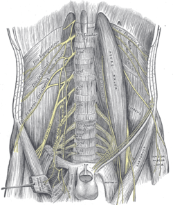 The lumbar plexus and its branches. (Femoral labeled at bottom left.) | |
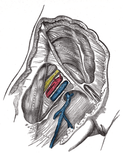 Femoral sheath laid open to show its three compartments. (Femoral nerve visible in yellow.) | |
| Details | |
| From | L2 |
| To | L4 |
| Innervates | anterior compartment of thigh |
| Identifiers | |
| Latin | nervus femoralis |
| MeSH | D005267 |
| TA | A14.2.07.020 |
| FMA | 16486 |
| Anatomical terms of neuroanatomy | |
Structure
Femoral nerve is the major nerve supplying the anterior compartment of the thigh. It is the largest branch of the lumbar plexus, and arises from the dorsal divisions of the ventral rami of the second, third, and fourth lumbar nerves (L2, L3, and L4).[1]
The nerve enters Scarpa's triangle by passing beneath the inguinal ligament, just lateral to the femoral artery. In the thigh, the nerve lies in a groove between iliacus muscle and psoas major muscles, outside the femoral sheath, and lateral to the femoral artery. After a short course of about 4 cm in the thigh, the nerve is divided into anterior and posterior divisions, separated by lateral femoral circumflex artery. The branches are shown below:[1]
Muscular branches
- The nerve to the Pectineus arises immediately above the inguinal ligament from the medial side of the femoral nerve, and passes behind the femoral sheath to enter the anterior surface of the muscle.[1]
- Anterior division supplies the Sartorius muscle[1]
- Posterior division supplies the rectus femoris, the three vasti (vastus medialis, vastus lateralis, and vastus intermedius), and articularis genus. The articularis genus is supplied by a branch of the nerve to vastus intermedius.[1]
Cutaneous branches
- The anterior division gives off Anterior cutaneous branches: The anterior cutaneous branches are: the intermediate femoral cutaneous nerve and the medial femoral cutaneous nerve.[1]
- The posterior division gives off only one branch, which is the saphenous nerve.[1]
Articular branches
Vascular branches
- Branches to the femoral artery and its branches.[1]
Clinical significance
Signals from the femoral nerve and its branches can be blocked to interrupt transmission of pain signal from the innervation area, by performing a regional nerve blockade. Some of the nerve blocks that works by affecting the femoral nerve are; femoral nerve block, fascia iliaca block and 3-in-1 nerve block.
Additional images
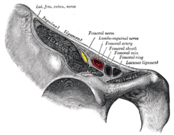 Structures passing behind the right inguinal ligament
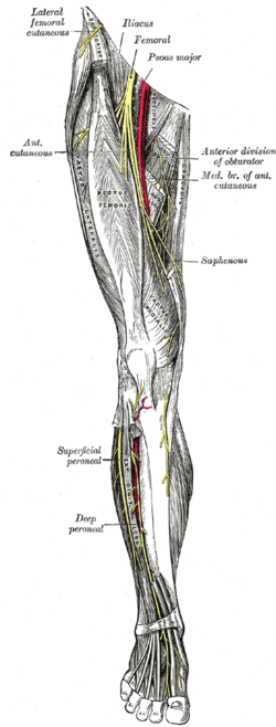 Nerves of the right leg.
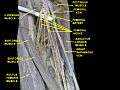 Femoral nerve.Deep dissection.
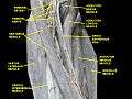 Femoral nerve.Deep dissection.
See also
References
This article incorporates text in the public domain from page 955 of the 20th edition of Gray's Anatomy (1918)
- Krishna, Garg (2010). "Front of the thigh (Chapter 3)". BD Chaurasia's Human Anatomy (Regional and Applied Dissection and Clinical) Volume 2 - Lower limb, abdomen, and pelvis (Fifth ed.). India: CBS Publishers and Distributors Pvt Ltd. p. 55. ISBN 978-81-239-1864-8.
External links
- Femoral_nerve at the Duke University Health System's Orthopedics program
- MedlinePlus Encyclopedia 000687 - "Femoral nerve dysfunction" (includes illustration)
- Anatomy photo:40:17-0202 at the SUNY Downstate Medical Center - "Posterior Abdominal Wall: Nerves of the Lumbar Plexus"
- Cross section image: pembody/body15a—Plastination Laboratory at the Medical University of Vienna
- Cross section image: pelvis/pelvis-e12-15—Plastination Laboratory at the Medical University of Vienna
- arteries-nerves%20LE/nerves1 at the Dartmouth Medical School's Department of Anatomy
| Authority control |
|---|