Fate mapping
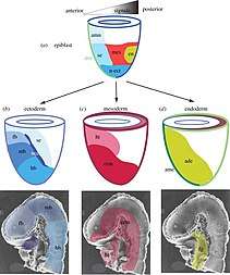
Fate mapping is a method used in developmental biology to study the embryonic origin of various adult tissues and structures. The "fate" of each cell or group of cells is mapped onto the embryo, showing which parts of the embryo will develop into which tissue. When carried out at single-cell resolution, this process is called cell lineage tracing.
History
The earliest fate maps were based on direct observation of the embryos of ascidians or other marine invertebrates.[1] Modern fate mapping began in 1929 when Walter Vogt marked the groups of cells using a dyed agar chip and tracked them through gastrulation.[2] In 1978, horseradish peroxidase (HRP) was introduced as a marker. HRP was more effective than previous markers, but required embryos to be fixed before viewing.[3] Genetic fate mapping is a technique developed in 1981 which uses a site-specific recombinase to track cell lineage genetically. Today, fate mapping is an important tool in many fields of biology research, such as developmental biology,[4] stem cell research, and kidney research.[5]
Cell Lineage
Fate mapping and cell lineage are similar but distinct topics, although there is often overlap. For example, the development of the complete cell lineage of C. elegans can be described as the fate maps of each cell division stacked hierarchically.[6] The distinction between the topics is in the type of information included. Fate mapping shows which tissues come from which part of the embryo at a certain stage in development, whereas cell lineage shows the relationships between cells at each division.[7] A cell lineage can be used to generate a fate map, and in cases like C. elegans, successive fate mapping is used to develop a cell lineage.[8]
See also
- Cell fate determination
References
- Conklin, Edwin Grant (1905). The organization and cell-lineage of the ascidian egg / by Edwin G. Conklin. Philadelphia: [Academy of Natural Sciences]. doi:10.5962/bhl.title.4801.
- Vogt, Walther (June 1929). "Gestaltungsanalyse am Amphibienkeim mit Örtlicher Vitalfärbung: II. Teil. Gastrulation und Mesodermbildung bei Urodelen und Anuren". Wilhelm Roux' Archiv für Entwicklungsmechanik der Organismen (in German). 120 (1): 384–706. doi:10.1007/BF02109667. ISSN 0949-944X. PMID 28354436.
- Weisblat, D.; Sawyer, R.; Stent, G. (1978-12-22). "Cell lineage analysis by intracellular injection of a tracer enzyme". Science. 202 (4374): 1295–1298. Bibcode:1978Sci...202.1295W. doi:10.1126/science.725606. ISSN 0036-8075.
- Gross, Joshua B.; Hanken, James (May 2008). "Review of fate-mapping studies of osteogenic cranial neural crest in vertebrates". Developmental Biology. 317 (2): 389–400. doi:10.1016/j.ydbio.2008.02.046. ISSN 0012-1606. PMID 18402934.
- Duffield, Jeremy S.; Humphreys, Benjamin D. (March 2011). "Origin of new cells in the adult kidney: results from genetic labeling techniques". Kidney International. 79 (5): 494–501. doi:10.1038/ki.2010.338. ISSN 0085-2538. PMID 20861816.
- Rudel, David; Sommer, Ralf J (December 2003). "The evolution of developmental mechanisms". Developmental Biology. 264 (1): 15–37. doi:10.1016/S0012-1606(03)00353-1.
- Hsu, Ya-Chieh (2015-08-18). "Theory and Practice of Lineage Tracing". Stem Cells. 33 (11): 3197–3204. doi:10.1002/stem.2123. ISSN 1066-5099. PMC 4618107.
- Liu, Xiao; Long, Fuhui; Peng, Hanchuan; Aerni, Sarah J.; Jiang, Min; Sánchez-Blanco, Adolfo; Murray, John I.; Preston, Elicia; Mericle, Barbara (October 2009). "Analysis of Cell Fate from Single-Cell Gene Expression Profiles in C. elegans". Cell. 139 (3): 623–633. doi:10.1016/j.cell.2009.08.044. ISSN 0092-8674. PMC 4709123. PMID 19879847.