Gestational sac
The gestational sac is the large cavity of fluid surrounding the embryo. During early embryogenesis it consists of the extraembryonic coelom, also called the chorionic cavity. The gestational sac is normally contained within the uterus. It is the only available structure that can be used to determine if an intrauterine pregnancy exists until the embryo is identified.
| Gestational sac | |
|---|---|
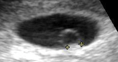 Contents in the cavity of the uterus seen at approximately 5 weeks of gestational age by obstetric ultrasonography. | |
 | |
| Details | |
| Carnegie stage | 6a |
| Days | 12 |
| Precursor | Heuser's membrane |
| Identifiers | |
| Latin | saccus gestationalis, coeloma extraembryonicum, cavitas chorionica |
| MeSH | D058746 |
| TE | E5.8.0.0.1.0.1 |
| Anatomical terminology | |
On obstetric ultrasound, the gestational sac is a dark ("anechoic") space surrounded by a white ("hyperechoic") rim.
Structure
The gestational sac is spherical in shape, and usually located in the upper part of the fundus of the uterus. By approximately 9 weeks of gestational age, the amniotic sac has expanded to occupy the majority of the volume of the gestational sac, eventually expanding to reduce the extraembryonic coelom to a thin layer between the amnion membrane and the mesoderm. By then, the gestational sac is usually simply called the "amniotic sac".
Development
During embryogenesis, the extraembryonic coelom (or chorionic cavity) that constitutes the gestational sac is a portion of the conceptus consisting of a cavity between Heuser's membrane and the Trophoblast.
During formation of the primitive yolk sac, some of the migrating hypoblast cells differentiate into mesenchymal cells that fill the space between Heuser's membrane and the Trophoblast, forming the extraembryonic mesoderm. As development progresses, small lacunae begin to form within the extraembryonic mesoderm which enlarge to become the extraembryonic coelom.
The extraembryonic coelom divides the extraembryonic mesoderm into two layers: extraembryonic splanchnopleuric mesoderm, which lies adjacent to Heuser's membrane around the outside of the primitive yolk sac, and extraembryonic somatopleuric mesoderm, which lies adjacent to the cytotrophoblast layer of the embryo.
The chorionic cavity is enclosed by the chorionic plate, which is composed of an inner layer of somatopleuric mesoderm and an outer layer of trophoblast cells.
Clinical significance
Ultrasound
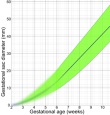
The mean sac diameter (MSD) can effectively estimate gestational age[2] between 5 and 6 weeks, with an accuracy of about +/- 5 days.[3]
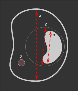
The yolk sac and embryo should be readily identifiable when the gestational sac reaches a certain size — a yolk sac should be seen when the gestational sac is 20mm and a fetal pole should be seen when the gestational sac reaches 25mm.
Gestational sacs can be identified via ultrasound and are generally identified by the following 4 characteristics:
- The sac has a round or elliptical shape in longitudinal and transverse views
- it is surrounded by a white echogenic rim ("choriodecidual reaction")
- The sac is located in the uterine fundus
- The sac is not implanted on the midline, but eccentrically (to one side of the uterine cavity line).
References
- Papaioannou, George I.; Syngelaki, Argyro; Poon, Leona C.Y.; Ross, Jackie A.; Nicolaides, Kypros H. (2010). "Normal Ranges of Embryonic Length, Embryonic Heart Rate, Gestational Sac Diameter and Yolk Sac Diameter at 6–10 Weeks". Fetal Diagnosis and Therapy. 28 (4): 207–219. doi:10.1159/000319589. ISSN 1421-9964. PMID 20847544.
Crown rump length before a gestational age of 6 weeks is extrapolated by assuming a gestational sac diameter of 0 at a gestational age of 2 weeks, corresponding to a fertilization age of 0 weeks. This extrapolated part is blurred because of uncertain values. - Karki DB, Sharmqa UK, Rauniyar RK (2006). "Study of accuracy of commonly used fetal parameters for estimation of gestational age". Journal of the Nepal Medical Association. 45 (162): 233–7. PMID 17189967.
- "Basic Imaging > Ultrasound of Early Pregnancy". Archived from the original on 2007-08-14. Retrieved 2007-10-13.
| Wikimedia Commons has media related to Gestational_sac. |
| Authority control |
|
|---|