Extensor retinaculum of the hand
The extensor retinaculum (dorsal carpal ligament, or posterior annular ligament) is an anatomical term for the thickened part of the antebrachial fascia that holds the tendons of the extensor muscles in place. It is located on the back of the forearm, just proximal to the hand. It is continuous with the palmar carpal ligament, which is located on the anterior side of the forearm.
| Extensor retinaculum of the hand | |
|---|---|
 | |
| Details | |
| Identifiers | |
| Latin | retinaculum musculorum extensorum manus |
| TA | A04.6.03.010 |
| FMA | 39987 |
| Anatomical terminology | |
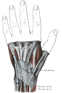
It is a strong, fibrous band, extending obliquely downward and medialward across the back of the wrist, and consisting of part of the deep fascia of the back of the forearm, strengthened by the addition of some transverse fibers.
The extensor retinaculum is attached laterally to the lateral margin of the radius. However, it is not attached to the ulna, as the distance between these two bones varies with supination and pronation of the forearm. Instead the medial attachment is to the most medial of the carpal bones, the triquetrum (or triquetral bone) and pisiformis (or pisiform bone). The retinaculum is also attached in its passage across the wrist, to the ridges on the dorsal surface of the radius.
Histology of the retinaculum
Structurally, the retinaculum consists of three layers. The deepest layer, the gliding layer, consists of hyaluronic acid-secreting cells. In studies of cadaver specimens this layer was found to show isolated chondroid metaplasia, an abnormal change in tissue which leads to it resembling cartilage. The thick middle layer consists of interspersed elastin fibers, collagen bundles, and fibroblasts. The most superficial layer is made up of loose connective tissue which contains vascular channels. Combined these three layers create a smooth gliding surface as well as mechanically strong tissue which prevents tendon bowstringing. The extensor retinaculum of the foot has similar structure.
Surgical uses
Studies conducted on the retinaculum have exhibited it to have several possible surgical treatments uses. A graft of the extensor retinaculum was shown to be useful in treating boxer's knuckle when direct repair of the damaged capsule is not possible. Because of their similarities in histological structure, studies also show the extensor retinaculum to be a reasonable biological replacement for reconstruction of a deficient annular pulley.
Additional images
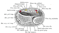 Transverse section across distal ends of radius and ulna.
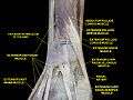 Extensor retinaculum of the hand.Deep dissection.
References
- Klein, David M., et al. "Histology of the extensor retinaculum of the wrist and the ankle." The Journal of Hand Surgery 24.4 (1999): 799-802.
- Nagaoka, Masahiro, et al. "Extensor retinaculum graft for chronic boxer’s knuckle." The Journal of Hand Surgery 31.6 (2006): 947-951.
External links
| Authority control |
|---|