Eosinophilic
Eosinophilic (Greek suffix -phil-, meaning loves eosin) refers to the staining of certain tissues, cells, or organelles after they have been washed with eosin, a dye.
Eosin is an acidic dye; thus, the structure being stained is basic and as a corollary, is acidophilic.
Eosinophilic describes the appearance of cells and structures seen in histological sections that take up the staining dye eosin. This is a bright-pink dye that stains the cytoplasm of cells, as well as extracellular proteins such as collagen.[1]
Such eosinophilic structures are, in general, composed of protein.
The stain eosin is usually combined with a stain called hematoxylin to produce a hematoxylin- and eosin-stained section (also called an H&E stain, HE or H+E section). This is the most widely used histological stain in medical diagnosis; for example, when a pathologist examines a biopsy of a suspected cancer, the biopsy will have been stained with H&E.
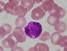
Some structures seen inside cells are described as being eosinophilic, for example, Lewy and Mallory bodies.[2] Some cells are also described as eosinophilic, such as Leukocytes.[3]
See also
- Basophilic (affinity to hematoxylin)
- Eosinophilia
- Eosinophilic meningitis
References
- About Eosinophilic Disorders. Children's Hospital of Philadelphia. Accessed March 2nd, 2012
- Eosinophilic. Medline Plus. Accessed March 2nd, 2012.
- Dixon, Frank J. (1986). Advances in Immunology, Volume 39. Academic Press. p. 323. ISBN 9780120224395.