Enthesitis
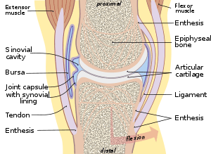
Enthesitis is inflammation of the entheses, the sites where tendons or ligaments insert into the bone.[1][2] It is an enthesopathy, a pathologic condition of the entheses. Early clinical manifestations are an aching sensation akin to "working out too much", and it gets better with activity. It is worst in the morning (after sleeping and not moving). The muscle insertion hurts very focally as it joins into the bone, but there is little to no pain at all with passive motion. While it is typically associated with other auto-immune diseases like spondyloarthropaties and psoriasis (thought to often precede psoriatic arthritis). A common autoimmune enthesitis is at the heel, where the Achilles tendon attaches to the calcaneus. There are some cases of isolated, primary enthesitis which are very poorly studied and understood.
| Enthesitis | |
|---|---|
 | |
| Typical joint showing the entheses | |
| Specialty | Rheumatology |
It is associated with HLA B27 arthropathies such as ankylosing spondylitis, psoriatic arthritis, and reactive arthritis.[3][4] Symptoms include multiple points of tenderness at the heel, tibial tuberosity, iliac crest, and other tendon insertion sites.
Images

Related conditions
Anatomically close but separate conditions are:
- Apophysitis, inflammation of the bony attachment, generally associated with overuse among growing children.[5][6][7]
- Tendinopathy is a disorder of the tendon, and is associated with direct injury or repetitive activities.[8]
See also
- Enthesis (plural: Entheses)
References
- Maria Antonietta D'Agostino, MD; Ignazio Olivieri, MD (June 2006). "Enthesitis". Best Practice & Research Clinical Rheumatology. Clinical Rheumatology. 20 (3): 473–86. doi:10.1016/j.berh.2006.03.007.
- The Free Dictionary (2009). "Enthesitis". Retrieved 2010-11-27.
- Schett, G; Lories, RJ; D'Agostino, MA; Elewaut, D; Kirkham, B; Soriano, ER; McGonagle, D (November 2017). "Enthesitis: from pathophysiology to treatment". Nature Reviews Rheumatology (Review). 13 (12): 731–41. doi:10.1038/nrrheum.2017.188. PMID 29158573.
- Schmitt, SK (June 2017). "Reactive Arthritis". Infectious Disease Clinics of North America (Review). 31 (2): 265–77. doi:10.1016/j.idc.2017.01.002. PMID 28292540.
- "OrthoKids - Osgood-Schlatter's Disease".
- "Sever's Disease". Kidshealth.org. Retrieved 2014-04-29.
- Hendrix CL (2005). "Calcaneal apophysitis (Sever disease)". Clinics in Podiatric Medicine and Surgery. 22 (1): 55–62, vi. doi:10.1016/j.cpm.2004.08.011. PMID 15555843.
- "Tendinitis". National Institute of Arthritis and Musculoskeletal and Skin Diseases. 12 April 2017. Retrieved 18 November 2018.