Cardiomegaly
Cardiomegaly is a medical condition in which the heart is enlarged. It is more commonly referred to as an enlarged heart. Other common names for cardiomegaly is megacardia or megalocardia, but they all refer to the same thing. The causes of cardiomegaly vary from patient to patient, depending on each case. Many times this condition results from high blood pressure (hypertension) or coronary artery disease. An enlarged heart may not pump blood effectively, resulting in congestive heart failure. Cardiomegaly may improve over time, but many people with an enlarged heart need lifelong treatment with medications.[12] Having an immediate family member who has or had cardiomegaly may indicate that a person is more susceptible to getting this condition.[13] Cardiomegaly is not a disease but rather a condition that can result from a host of other diseases such as obesity or coronary artery disease. Recent studies suggest that cardiomegaly is associated with a higher risk of sudden cardiac death (SCD).[14] Cardiomegaly can be serious even though it is not an actual disease. Depending on what part of the heart is enlarged, the patient can suffer from a heart failure. Anyone can experience a heart enlargement, but not everyone will be diagnosed immediately, based on the signs and symptoms. Cardiomegaly leads to clinical heart failure and in United States nearly 5.8 million people suffer. Heart failures increase with age, more common in males, and African Americans. According to research conducted in June 2019, mentioned that half of the people diagnosed with heart failure die within 5 years, after the diagnosis. Cardiomyopathy is also associated with cardiomegaly, and it is a disease of the heart muscle, which makes it difficult for the heart to pump blood to the rest of the body. Cardiomyopathy can lead to a heart failure. There are three main types of cardiomyopathy: hypertrophic, dilated, and restrictive. The difference is that cardiomegaly is a condition of the heart and cardiomyopathy is an actual disease.
| Cardiomegaly | |
|---|---|
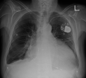 | |
| Cardiomegaly on chest X-ray with a pacemaker | |
| Specialty | Cardiology |
| Types | Athletic heart syndrome,[1] Ventricular hypertrophy, Atrial enlargement |
| Causes | Dilated cardiomyopathy,[2][3][4][5] Hypertrophic cardiomyopathy.[1][6][7][8][9] |
| Diagnostic method | Hypertrophic cardiomyopathy screening[10][11] |
Signs and symptoms
For many people, cardiomegaly is asymptomatic. For others, if the enlarged heart begins to affect the body's ability to pump blood effectively, then symptoms associated with congestive heart failure may arise, including:[13]
- Heart palpitations – the irregular beating of the heart, usually associated with a valve issue inside the heart.
- Severe shortness of breath (especially when physically active) – irregularly unable to catch one's breath.
- Chest pain
- Coughing, when lying down
- Fatigue
- Swelling in legs
- Increased abdominal girth
- Weight gain
- Edema – swelling
- Fainting[13]
There is not much variation in these symptoms because they are mostly specific to the chest area. However, some are more common than others depending on each patient.
Causes & Prevention
The causes of cardiomegaly are not well understood and many cases of cardiomegaly are idiopathic (having no known cause). Prevention of cardiomegaly starts with detection. If a person has a family history of cardiomegaly, one should let one's doctor know so that treatments can be implemented to help prevent worsening of the condition. In addition, prevention includes avoiding certain lifestyle risk factors such as tobacco use and controlling one's high cholesterol, high blood pressure, and diabetes. Non-lifestyle risk factors include family history of cardiomegaly, coronary artery disease (CAD), congenital heart failure, Atherosclerotic disease, valvular heart disease, exposure to cardiac toxins, sleep disordered breathing (such as sleep apnea), sustained cardiac arrhythmias, abnormal electrocardiograms, and cardiomegaly on chest X-ray. Lifestyle factors which can help prevent cardiomegaly include eating a healthy diet, controlling blood pressure, exercise, medications, and not abusing alcohol and cocaine.[13] Current research and the evidence of previous cases link the following (below) as possible causes of cardiomegaly.
The most common causes of cardiomegaly are congenital (patients are born with the condition based on a genetic inheritance), high blood pressure which can enlarge the left ventricle causing the heart muscle to weaken over time, and coronary artery disease that creates blockages in the heart's blood supply, which can bring on a cardiac infarction (heart attack) leading to tissue death which causes other areas of the heart to work harder, increasing the heart's size.
Other possible causes include:
- Heart Valve Disease
- Cardiomyopathy (disease of the heart muscle)
- Pulmonary Hypertension
- Pericardial Effusion (fluid around the heart)
- Thyroid Disorders
- Hemochromatosis (excessive iron in the blood)
- Other rare diseases like Amyloidosis[13]
- Chagas disease, an important cause of cardiomegaly in Latin America[15]
- Viral infection of the heart
- Pregnancy, with enlarged heart developing around the time of delivery (peripartum cardiomyopathy)
- Kidney disease requiring dialysis
- Alcohol or cocaine abuse
- HIV infection[12]
- Diabetes[16]
Mechanism
Cardiomegaly is a condition affecting the cardiovascular system, specifically the heart. This condition is strongly associated with congestive heart failure.[13] Within the heart, the working fibers of the myocardial tissue increase in size. As the heart works harder the actin and myosin filaments experience less overlap which increases the size of the myocardial fibers. If there is less overlap of the protein filaments actin and myosin within the sarcomeres of muscle fibers, they will not be able to effectively pull on one another. If the heart tissue (walls of left and right ventricle) gets too big and stretches too far, then those filaments cannot effectively pull on one another to shorten the muscle fibers, thus impacting the heart's sliding filament mechanism. If fibers cannot shorten properly, and the heart cannot contract properly, then blood cannot be effectively pumped to the lungs to be re-oxygenated and to the body to deliver oxygen to the working tissues of the body.
Person with an enlarged heart is more susceptible to forming blood clots in the lining of their heart. These clots can also be formed in other parts of the body. Once they enter the bloodstream, it makes it difficult for the organs in the body to receive blood, due to the blockage caused by the clots. This can impact other body systems as well and lead to other problems.
Diagnosis
There are many techniques and tests used to diagnose an enlarged heart. The results of these tests can often be used to see how efficiently the heart is pumping, determine which chambers of the heart are enlarged, look for evidence of previous heart attacks and determine if a person has congenital heart disease.
Risk factors for cardiomegaly include: a family history of heart disease, diabetes, obesity, hypertension, history of alcohol or drug abuse, lifestyle that consists of little or no exercise.
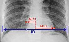

where:[17]
MRD = greatest perpendicular diameter from midline to right heart border
MLD = greatest perpendicular diameter from midline to left heart border
ID = internal diameter of chest at level of right hemidiaphragm
- Chest X-Ray: X-ray images help see the condition of the lungs and heart. If the heart is enlarged on an X-ray, other tests will usually be needed to find the cause. A useful measurement on X-ray is the cardio-thoracic ratio, which is the transverse diameter of the heart, compared with that of the thoracic cage."[18] These diameters are taken from PA chest x-rays using the widest point of the chest and measuring as far as the lung pleura, not the lateral skin margins. If the cardiac thoracic ratio is greater than 50%, pathology is suspected, assuming the x-ray has been taken correctly.[19] The measurement was first proposed in 1919 to screen military recruits. A newer approach to using these x-rays for evaluating heart health takes the ratio of heart area to chest area and has been called the two-dimensional cardiothoracic ratio.[20]
- Electrocardiogram: This test records the electrical activity of the heart through electrodes attached to the person's skin. Impulses are recorded as waves and displayed on a monitor or printed on paper. This test helps diagnose heart rhythm problems and assess the damage to a person's heart from a heart attack.
- Echocardiogram: This test for diagnosing and monitoring an enlarged heart uses sound waves to produce a video image of the heart. With this test, the four chambers of the heart can be evaluated.
- Stress test: A stress test, also called an exercise stress test, provides information about how well the heart works during physical activity. It usually involves walking on a treadmill or riding a stationary bike while the heart rhythm, blood pressure, and breathing are monitored.
- Cardiac computerized tomography (CT) or magnetic resonance imaging (MRI). In a cardiac CT scan, one lies on a table inside a machine called a gantry. An X-ray tube inside the machine rotates around the body and collects images of the heart and chest. In a cardiac MRI, one lies on a table inside a long tube-like machine that uses a magnetic field and radio waves to produce signals that create images of the heart.
- Blood tests: Blood tests may be ordered to check the levels of substances in the blood that may show a heart problem. Blood tests can also help rule out other conditions that may cause one's symptoms.
- Cardiac catheterization and biopsy: In this procedure, a thin tube (catheter) is inserted in the groin and threaded through the blood vessels to the heart, where a small sample (biopsy) of the heart, if indicated, can be extracted for laboratory analysis.[13]
Classification
Cardiomegaly can be classified by the main enlarged location of the heart, and/or by the structure of the enlargement.
There are also specific additional subtypes. For example, the athletic heart syndrome is a non-pathological condition commonly seen in sports medicine in which the human heart is enlarged, and the resting heart rate is lower than normal.
By enlarged location
Structure of enlargement
Dilated cardiomyopathy is the most common type of cardiomegaly. In this condition, the walls of the left and/or right ventricles of the heart become thin and stretched. The result is an enlarged heart.
In the other types of cardiomegaly, the heart's large muscular left ventricle becomes abnormally thick. Hypertrophy is usually what causes left ventricular enlargement. Hypertrophic cardiomyopathy is typically an inherited condition.[12]
Treatment and prognosis
Treatments for cardiomegaly include a combination of medication treatment and medical/surgical procedures. Below are some of the treatment options for individuals with cardiomegaly:
Medications
- Diuretics: to lower the amount of sodium and water in the body, which can help lower the pressure in the arteries and heart.
- Angiotensin-converting enzyme (ACE) inhibitors: to lower the blood pressure and improve the heart's pumping ability.
- Angiotensin receptor blockers (ARBs): to provide the benefits of ACE inhibitors for those who can't take ACE inhibitors.
- Beta blockers: to lower blood pressure and improve heart function.
- Digoxin: to help improve the pumping function of the heart and lessen the need for hospitalization for heart failure.
- Anticoagulants: to reduce the risk of blood clots that could cause a heart attack or stroke.
- Anti-arrhythmics: to keep the heart beating with a normal rhythm.
Medical devices to regulate the heartbeat
- Pacemaker: Coordinates the contractions between the left and right ventricle. In people who may be at risk of serious arrhythmias, drug therapy or an implantable cardioverter-defibrillator (ICD) may be used.
- ICDs: Small devices implanted in the chest to constantly monitor the heart rhythm and deliver electrical shocks when needed to control abnormal, rapid heartbeats. The devices can also work as pacemakers.
Surgical procedures
- Heart valve surgery: If an enlarged heart is caused by a problem with one of the heart valves, one may have surgery to remove the valve and replace it with either an artificial valve or a tissue valve from a pig, cow or deceased human donor. If blood leaks backward through a valve (valve regurgitation), the leaky valve may be surgically repaired or replaced.
- Coronary bypass surgery: If an enlarged heart is related to coronary artery disease, one may opt to have coronary artery bypass surgery.
- Left ventricular assist device: (LVAD): This implantable mechanical pump helps a weak heart pump. LVADs are often implanted while a patient waits for a heart transplant or, if the patient is not a heart transplant candidate, as a long-term treatment for heart failure.
- Heart transplant: If medications can't control the symptoms, a heart transplant is often a final option.[13]
Cardiomegaly can progress and certain complications are common:
- Heart failure: One of the most serious types of enlarged heart, an enlarged left ventricle, increases the risk of heart failure. In heart failure, the heart muscle weakens, and the ventricles stretch (dilate) to the point that the heart can't pump blood efficiently throughout the body.
- Blood clots: Having an enlarged heart may make one more susceptible to forming blood clots in the lining of the heart. If clots enter the bloodstream, they can block blood flow to vital organs, even causing a heart attack or stroke. Clots that develop on the right side of the heart may travel to the lungs, a dangerous condition called a pulmonary embolism.
- Heart murmur: For people who have an enlarged heart, two of the heart's four valves — the mitral and tricuspid valves — may not close properly because they become dilated, leading to a backflow of blood. This flow creates sounds called heart murmurs.
- The exact mortality rate for people with cardiomegaly is unknown. However, many people live for a very long time with an enlarged heart and if detected early, treatment can help improve the condition and prolong the lives of these people.[13]
- For some people cardiomegaly is a temporary condition, which can resolve on its own, making one's lifestyle normal like before. Others may have a permanent enlargement, which would then need to be taken care of by the above treatment options.
At home remedies
- Stop smoking
- Limit alcohol and caffeine intake
- Maintain healthy weight
- Increasing fruits and vegetables in daily diet
- Cutting down on high fat foods and high sugar foods
- Monitoring blood pressure
- Sleeping 7-9 hours every night
Recent Research
In a recent study published in 2000, researchers focus on the impact cardiomegaly has on lung dysfunction in patients with heart failure. The hypothesis they made was proven to be correct, after the study conducted in New York came in. An enlarged heart competes for space with the lungs, hence it causes a restrictive lung pattern and reduces alveolar volume. Another study conducted in Germany mentions the efforts made to understand the molecular signaling pathways mediating cardiac growth. Understanding the pathway can aid in new drug treatment options for the new generation.
Another interesting study that was done in 2018 proved a common idea wrong. People tend to believe that the size of heart is equivalent to the size of the person's fist. However, after doing some research the article suggested that the idea might actually be incorrect. This assumptions leads to misdiagnosis during autopsy, but further study is still being done to prove the idea.
References
- "Enlarged heart". Heart and Stroke Foundation of Canada. Retrieved 2019-03-29.
Types...Hypertrophic cardiomyopathy, Left ventricular hypertrophy (LVH), Intense, prolonged athletic training
- Hershberger, Ray E; Morales, Ana; Siegfried, Jill D (22 September 2010). "Clinical and genetic issues in dilated cardiomyopathy: A review for genetics professionals". Genetics in Medicine. 12 (11): 655–667. doi:10.1097/GIM.0b013e3181f2481f. PMC 3118426. PMID 20864896.
- Luk, A; Ahn, E; Soor, G S; Butany, J (18 November 2008). "Dilated cardiomyopathy: a review". Journal of Clinical Pathology. 62 (3): 219–225. doi:10.1136/jcp.2008.060731. PMID 19017683.
- "What Is an Enlarged Heart (Cardiomegaly)?". WebMD. 2019-01-30. Retrieved 2019-03-29.
- Lee, Ji Eun; Oh, Jin-Hee; Lee, Jae Young; Koh, Dae Kyun (2014). "Massive Cardiomegaly due to Dilated Cardiomyopathy Causing Bronchial Obstruction in an Infant". Journal of Cardiovascular Ultrasound. 22 (2): 84–7. doi:10.4250/jcu.2014.22.2.84. PMC 4096670. PMID 25031799.
- Marian, Ali J.; Braunwald, Eugene (15 September 2017). "Hypertrophic Cardiomyopathy". Circulation Research. 121 (7): 749–770. doi:10.1161/CIRCRESAHA.117.311059. PMC 5654557. PMID 28912181.
- Maron, Martin S (1 February 2012). "Clinical Utility of Cardiovascular Magnetic Resonance in Hypertrophic Cardiomyopathy". Journal of Cardiovascular Magnetic Resonance. 14 (1): 13. doi:10.1186/1532-429X-14-13. PMC 3293092. PMID 22296938.
- Almog, C; Weissberg, D; Herczeg, E; Pajewski, M (1 February 1977). "Thymolipoma simulating cardiomegaly: a clinicopathological rarity". Thorax. 32 (1): 116–120. doi:10.1136/thx.32.1.116. PMC 470537. PMID 138960.
- Hou, Jianglong; Kang, Y. James (September 2012). "Regression of pathological cardiac hypertrophy: Signaling pathways and therapeutic targets". Pharmacology & Therapeutics. 135 (3): 337–354. doi:10.1016/j.pharmthera.2012.06.006. PMC 3458709. PMID 22750195.
- Luis Fuentes, Virginia; Wilkie, Lois J. (September 2017). "Asymptomatic Hypertrophic Cardiomyopathy". Veterinary Clinics of North America: Small Animal Practice. 47 (5): 1041–1054. doi:10.1016/j.cvsm.2017.05.002. PMID 28662873.
- Maron, Barry J; Maron, Martin S (January 2013). "Hypertrophic cardiomyopathy". The Lancet. 381 (9862): 242–255. doi:10.1016/S0140-6736(12)60397-3. PMID 22874472.
- "What Is an Enlarged Heart (Cardiomegaly)?". WebMD.
- "Enlarged heart - Symptoms and causes". mayoclinic.org. Retrieved 19 March 2018.
- Tavora F; et al. (2012). "Cardiomegaly is a common arrhythmogenic substrate in adult sudden cardiac deaths, and is associated with obesity". Pathology. 44 (3): 187–91. doi:10.1097/PAT.0b013e3283513f54. PMID 22406485.
- Bestetti, Reinaldo B. (Nov 2016). "Chagas Heart Failure in Patients from Latin America". Card Fail Rev. 2 (2): 90–94. doi:10.15420/cfr.2016:14:2. PMC 5490952. PMID 28785459.
- http://www.ddcmultimedia.com/doqit/Care_Management/CM_HeartFailure/L1P4.html%5B%5D
- "Chest Measurements". Oregon Health & Science University. Retrieved 2017-01-13.
- "cardiothoracic ratio". thefreedictionary.com. Retrieved 19 March 2018.
- Justin, M; Zaman, S; Sanders, J.; Crook, A. M; Feder, G.; Shipley, M.; Timmis, A.; Hemingway, H. (1 April 2007). "Cardiothoracic ratio within the 'normal' range independently predicts mortality in patients undergoing coronary angiography". Heart. 93 (4): 491–494. doi:10.1136/hrt.2006.101238. PMC 1861494. PMID 17164481.
- Browne, Ronan F. J.; O’Reilly, Geraldine; McInerney, David (18 February 2004). "Extraction of the Two-Dimensional Cardiothoracic Ratio from Digital PA Chest Radiographs: Correlation with Cardiac Function and the Traditional Cardiothoracic Ratio". Journal of Digital Imaging. 17 (2): 120–123. doi:10.1007/s10278-003-1900-3. PMC 3043971. PMID 15188777.
- Tracy, Richard Everett (2011). "Association of Cardiomegaly with Coronary Artery Histopathology and its Relationship to Atheroma". Journal of Atherosclerosis and Thrombosis. 18 (1): 32–41. doi:10.5551/jat.5090. PMID 20953090.
Further reading
- Amin, Hina; Siddiqui, Waqas J. (2019). "Cardiomegaly". StatPearls. StatPearls Publishing.
- Ampanozi, Garyfalia; Krinke, Eileen; Laberke, Patrick; Schweitzer, Wolf; Thali, Michael J.; Ebert, Lars C. (1 September 2018). "Comparing fist size to heart size is not a viable technique to assess cardiomegaly". Cardiovascular Pathology. 36: 1–5. doi:10.1016/j.carpath.2018.04.009.
- Agostoni, PierGiuseppe; Cattadori, Gaia; Guazzi, Marco; Palermo, Pietro; Bussotti, Maurizio; Marenzi, Giancarlo (1 November 2000). "Cardiomegaly as a possible cause of lung dysfunction in patients with heart failure". American Heart Journal. 140 (5): A17–A21. doi:10.1067/mhj.2000.110282. PMID 11054632.
- Luedde, Mark; Katus, Hugo; Frey, Norbert (1 January 2006). "Novel Molecular Targets in the Treatment of Cardiac Hypertrophy". Recent Patents on Cardiovascular Drug Discovery. 1 (1): 1–20. doi:10.2174/157489006775244290. PMID 18221071.
External links
| Classification |
|---|