Diff-Quik
Diff-Quik is a commercial Romanowsky stain variant used to rapidly stain and differentiate a variety of pathology specimens. It is most frequently used for blood films and cytopathological smears, including fine needle aspirates.[1][2][3] The Diff-Quik procedure is based on a modification of the Wright-Giemsa stain pioneered by Harleco in the 1970s,[1] and has advantages over the routine Wright-Giemsa staining technique in that it reduces the 4-minute process into a much shorter operation and allows for selective increased eosinophilic or basophilic staining depending upon the time the smear is left in the staining solutions.[4]
There are generic brands of such stain,[1][5] and the trade name is sometimes used loosely to refer to any such stain (much as "Coke" or "Band-Aid" are sometimes used imprecisely).
Usage
Diff-Quik may be utilized on material which is air-dried prior to alcohol fixation rather than immersed immediately (i.e. "wet-fixed"), although immediate alcohol fixation results in improved microscopic detail.[2][3][6]
The primary use of Romanowsky-type stains in cytopathology is for cytoplasmic detail, while Papanicolaou stain is used for nuclear detail. Diff-Quik stain highlights cytoplasmic elements such as mucins, fat droplets and neurosecretory granules. Extracellular substances, such as free mucin, colloid, and ground substance, are also easily stained, and appear metachromatic. Microbiologic agents, such as bacteria and fungi, also appear more easily in Diff-Quik.[3] The stain can be used to detect Helicobacter pylori,[5] and it may also be used in the evaluation of sperm morphology.[7]
Due to its short staining time, Diff-Quik stain is often used for initial screening of cytopathology specimens. This staining technique allows the cytotechnologist or pathologist to quickly assess the adequacy of the specimen, identify possible neoplastic or inflammatory changes, and decide whether or not additional staining is required.[4][8][9]
Components
The Diff-Quik stain consists of 3 solutions:[4]
- Diff-Quik fixative reagent
- Triarylmethane dye
- Methanol
- Diff-Quik solution I (eosinophilic)
- Xanthene dye (Eosin Y)
- pH buffer
- Diff-Quik solution II (basophilic)
- Thiazine dye, methylene blue and azure A
- pH buffer
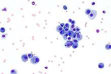
Results
| Structure | Colour |
|---|---|
| Erythrocytes | Pink/yellowish red |
| Platelets | Violet/purple granules |
| Neutrophils | Blue nucleus, pink cytoplasm, violet granules |
| Eosinophils | Blue nucleus, blue cytoplasm, red granules |
| Basophils | Purple/dark blue nucleus, violet granules |
| Monocyte | Violet nucleus, light blue cytoplasm |
| Bacteria and fungi | Dark blue |
| Cytoplasm, collagen and muscle | Various shades of pink, orange, yellow and blue[10] |
| Spermatozoa | Light blue acrosomal region, dark blue post-acrosomal region[7] |
Alternatives
- Wright Giemsa stain
- Papanicolaou stain
References
- Silverman JF, Frable WJ (1990). "The use of the diff-quik stain in the immediate interpretation of fine-needle aspiration biopsies". Diagn Cytopathol. 6 (5): 366–9. doi:10.1002/dc.2840060516. PMID 1705500.
- Susan C. Lester (10 May 2019). "Section 2: Methods - Slide preparation". Diagnostic Pathology: Intraoperative Consultation (2nd ed.). Elsevier Health Sciences. p. 69. ISBN 978-0-323-57020-6.
- Demay, Richard (2012). "Chapter 26: Stains". The art and science of cytopathology. Chicago, IL: Am Soc Clinical Pathology. p. 1505. ISBN 978-0-89189-644-9. OCLC 761848930.
- Keebler, Catherine (1993). The manual of cytotechnology (7th ed.). Chicago: ASCP Press. ISBN 0-89189-352-0. OCLC 27435280.
- Kumar, George L.; Kiernan, John A.; Gill, Gary W.; Badve, Sunil (2010). "Chapter 1: Introduction to special stains" (PDF). Education guide: Special stains and H&E (2nd ed.). Dako North America. p. 5. Archived (PDF) from the original on 2019-01-30. Retrieved 2019-06-29.
- William G. Finn; LoAnn C. Peterson (31 May 2004). "Role of fine needle aspiration in lymphoma". Hematopathology in Oncology. Springer Science & Business Media. p. 184. ISBN 978-1-4020-7919-1.
- "Part I: Semen analysis". WHO laboratory manual for the examination and processing of human semen (5th ed.). Geneva: World Health Organization. 2010. p. 62. ISBN 978-92-4-154778-9. OCLC 646393549.
- Meena, Nikhil; Jeffus, Susanne; Massoll, Nicole; Siegel, Eric R.; Korourian, Soheila; Chen, Chien; Bartter, Thaddeus (2015). "Rapid onsite evaluation: A comparison of cytopathologist and pulmonologist performance". Cancer Cytopathology. Wiley. 124 (4): 279–284. doi:10.1002/cncy.21637. ISSN 1934-662X. PMID 26492064.
- Roh, Michael H. (2015). "The Utilization of Cytologic Fine-Needle Aspirates of Lung Cancer for Molecular Diagnostic Testing". Journal of Pathology and Translational Medicine. The Korean Society of Pathologists and The Korean Society for Cytopathology. 49 (4): 300–309. doi:10.4132/jptm.2015.06.16. ISSN 2383-7837. PMC 4508567. PMID 26076721.
- Polysciences Inc. "Differential Quik Stain (Modified Giemsa)" (PDF). Technical Data Sheet #715. Archived (PDF) from the original on 2019-06-30. Retrieved 2019-06-30.