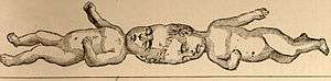
Craniopagus twins
Craniopagus twins are conjoined twins that are fused at the cranium.[1] Conjoined twins occur in about 10–20 babies in every million births in the United States. Among this small group, cephalic conjoining, or craniopagus twinning, represents the rarest of congenital abnormalities, accounting for 2–6% of all conjoined twins. Additionally, conjoined twins are genetically identical and always share the same sex. The union in craniopagus twins may occur on any portion of the cranium, but does not include either the face or the foramen magnum. The thorax and abdomen are separate and each twin has its own umbilicus and umbilical cord.
| Craniopagus twins | |
|---|---|
 | |
| Craniopagus twins | |
| Specialty | Medical genetics |
The union may involve the entire diameter of the head or only a small portion. This suggests that although many different kinds of vulnerabilities are already known in the scientific community, there are an infinite number of variations that can occur. Most of these variations are based on the rotation of one twin's skull to the other and the different phenotype sub-groups of craniopagus twins are based on all these rotational conformations. Each of these factors (rotation, spot of union) affects the development of the brain, the vascular system within the brain and overall wellness of life both of the twins have outside the womb.
Relatively few craniopagus twins survive the perinatal period – approximately 40% of conjoined twins are stillborn and an additional 33% die within the immediate perinatal period, usually from organ abnormalities and failure.[2] However 25% of craniopagus twins survive and can be considered for a surgical separation and several attempts occur yearly worldwide. In the last-half century, many advances in medicine including brain imaging, neuro-anesthesia and neurosurgical techniques have proven that a successful outcome is possible following separation of total craniopagus twins.
Categories
There are two categories of craniopagus twins:
- Partial: Partial craniopagus is less common than total. It is defined as having limited surface area involvement, with either intact crania or cranial defects. In other words, it is a defect of the cranial "coverings." In partial craniopagus twins, the unions are usually frontal and less commonly occipital and vertical. Angular frontal junctions occur when the two twins are joined at any part of the forehead. Occipital twins are joined at the occipital lobe in the back of the head and vertical are joined on the top of the head and usually face opposite directions. The junctional diameter is often smaller in partial forms and occasionally an incomplete layer of bone may be present between the twins. Each child maintains independent calvarial convexities except at the common area of skull junction. The dura of both children may be intact or deficient and cortical gyri may interdigitate.[3] Additionally shared dural venous sinuses is usually absent or if it is present it is negligible. These twins usually undergo successful separation and both twins may live to lead normal lives.
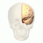
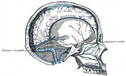
- Total: Total craniopagus twins are defined as sharing extensive surface area with widely connected cranial cavities. Among total craniopagus twins, there are four main categories which are then further divided into several subcategories. Frontal, the first category, are when twins are facing each other with the axis of the bodies forming an acute angle. Temporoparietal craniopagi are joined immediately above the external auditory meatus.[4] The third division is the occipital anomaly where the twins are connected in the occipital lobe causing the twins to face away from each other. The final variant is the parietal craniopagus which occurs when twins fuse at the vertex with the axis of the twins forming an obtuse angle. This category is perhaps the most important, or most interesting because the craniums of the two twins share the most veins, lobes and circuitry and is often described as one brain shared by two individuals.
- Having this kind of juncture means that there would be one common continuous cranium housing four cerebral hemispheres. An incomplete dural septum typically separates the flattened cerebral hemispheres. In total vertical craniopagus, the major cerebral arterial supply is usually confined to each respective twin and in some cases conjoined brain tissue may contain a larger artery. Within this category there are three smaller subdivisions that basically outline the different rotational symmetry of the junction[5]
- Type 1: both children face in the same general directional axis so that the angle between twins is less than 40 degrees. These twins show relatively symmetric superior bi-parietal or vertex compressional flattening.
- Type 2: both children face opposite directions so that the deformity shows an axial rotation between 140-180 degrees.
- Type 3: in this variety axial rotation is intermediate between the first two types with a rotation of being between 40 and 140.
Gestation and embryology
The exact nature of how conjoined twins develop inutero remains unclear. Embryologists have traditionally attributed identical twinning as "splitting or fission" of either the inner cell mass of pleuripotential cells or early embryonic disc at 13–14 days of gestation just before the primitive streak. Some theorists suggested that conjoined twins develop as a result of the failed fusion of a single fertilized ovum. However a new hypothesis suggests that cranial fusion occurs between two separate embryos before the end of the 4th week of gestation. This can happen because the cranial neuropore is still open, which is responsible for the ultimate fusion and formation of the brain stem and central nervous system. Furthermore, this secondary fusion of embryonic discs could implicate that intact skin will not fuse to other intact skin, including the ectoderm of the embryo. This means that two embryonic discs could only unite in locations where the ectoderm is absent. Moreover, the fusion occurs from neural folds of two separate, dorsally oriented embryonic discs, and the union can occur only after the ectoderm is disrupted to allow the neural and surface ectodermal layers to separate from each other. The union in craniopagus twins may happen at any portion of the calvarium.[6] The juncture can involve either the entire diameter of the head or any portion of the head and can be positioned at a multitude of rotational angles. In fact, craniopagus twins are rarely found in a symmetrical union. Apart from this, the vertebral axes may have a straight line. Despite this, the angle of the vertebrae is the ultimate dictator in how the individuals heads actually face. The majority of twins face either the same way or the exact opposite direction. Many reviews suggest a practical four-category system that breaks down the craniopagus twins on the basis of vertical or angular configuration or on the basis if there were shared dural venous sinuses. This scheme was applied to 64 cases and has adequately described sets of twins for over the last 86 years.
Further phenotypic divisions of cranial unions
- Vertical craniopagus: twins are joined at the top of the head with bodies at 180 degree angles to each other.[7]
- Occipital craniopagus: twins are joined at the back of the head
- Frontal craniopagus: twins are joined at the forehead.
- Parietal craniopagus: twins are joined at the side of the head
Medical procedures
- Conjoined twins can be diagnosed using standard ultrasound equipment during mid-pregnancy.
- Treatment of conjoined twins varies largely depending on the type of union and the circumstances of the twins.
- Many parents make the decision to terminate pregnancy due to the prognosis and quality of life issues that the twins would have to deal with. Also the likelihood of a successful separation can vary a parents likelihood of terminating pregnancy.
- If the parents choose to continue the pregnancy, mother and babies will be closely monitored throughout the pregnancy. In almost all cases a surgical procedure (C-section) delivery is planned often two to three weeks before the due date.[8]
- After the twins are born, parents and doctors decide whether or not separation surgery is possible weighing in many things such as if the twins share vital organs, are the twins healthy enough to withstand surgery and how good the odds are. The doctors also must consider the possibility of reconstructive surgery and the social and learning issues the twins may have to face after they are separated.
Cases
- Emilie and Elisabeth Stoll were born January 17, 1912. Their parents exhibited them until their death in July 1912. Their cause of death is unknown, but they were probably weakened by the stress of travelling and exhibition.[9]
- Rodney and Roger Brodie were born in Rock Island, Illinois in 1951. In December, 1952 a medical team led by neurosurgeon Dr. Oscar Sugar attempted to separate the fifteen-month old twins in a surgery that lasted twelve hours. During surgery, the doctors discovered that the twins shared the sagittal sinus, the canal that drains blood from the brain to the heart. This vessel was retained by Rodney Brodie. Both twins survived the surgery, although Roger Brodie did not regain consciousness and died 34 days later. Rodney Brodie recovered from the surgery but did suffer from neurological damage. Because his skull was never closed he wore a helmet until his death at age 11. This was the first case where craniopagual twins were separated and one survived.[10]
- Lotti and Rosemarie Knaack, who were born in Germany in 1951. They were separated at age 6, with Lotti dying in surgery. Rosemarie survived until 2008.
- Lori and George Schappell, born 1961, are believed to be the longest-surviving craniopagus twins still living.
- Ladan and Laleh Bijani, who were separated at age 29, only to die 90 minutes apart after surgery.
- Ahmed and Mohamed Ibrahim who were born in Egypt. They were successfully separated at age 2 years, 4 months in October 2001 in Dallas, Texas.[11]
- Lea and Tabea Block, who were born in Germany in 2003. Their separation surgeries were in September 2004 at Johns Hopkins Hospital in Baltimore, Maryland. Tabea died of cardiac arrest an hour after separation.
- Anastasia and Tatiana Dogaru, who were born with the crown of Tatiana's head joined to the back of Anastasia's. Doctors determined in 2007 that they could not be separated.[12]
- Krista and Tatiana Hogan, born on October 25, 2006. After a series of tests doctors also determined these twins could also not be separated.
- Joseph and Luka Banda from Zambia, born 1997, were separated successfully that year in South Africa by a team of surgeons led by Ben Carson.
- Trishna and Krishna from Bangladesh born in December 2006, joined on the tops of their skulls and sharing a small amount of brain tissue. In 2009, they were separated in Melbourne, Australia.[13]
- Ganga and Jamuna Shreshta, born in Kathmandu, Nepal, successfully separated by Dr. Chumpon Chan and his team from the Singapore General Hospital.
- Rital and Ritaj Gaboura, born in Sudan, were separated at 11 months old in 2011 at the Great Ormond Street Hospital in London. As of 2019 they lived in Ireland.[14]
- Jadon and Anias McDonald were separated at 13-months in 2016 in a crowdfunded operation costing $2.5million (USD) at Montefiore Hospital in New York.[15]
- Maria Ysadora and Maria Ysabelle, born in July, 2016. In October 27, 2018, they were successfully separated after a multistage surgery (five steps) in Ribeirao Preto, Sao Paulo - Brazil by a multidisciplinary team. Ricardo Santos de Oliveira (neurosurgeon) and Marcelo Volpon (neurosurgeon), Jayme Farina and Pedro Soler (plastic surgery). The team was led by Helio Machado and James Tait Goodrich. The surgery was done in University Hospital from Ribeirao Preto School of Medicine, University of Sao Paulo.
- Safa and Marwa Bibi, born in Peshawar, Pakistan on January 7, 2017, were separated in February 2019 during the third of three surgeries.[14]
- Islam Twins: Rabeya and Rukaya Islam, born in Pabna, Bangladesh on July 16, 2016. As the 3rd stage of their separation surgery series called “Operation Freedom, they were successfully separated after a 33-hour surgery in Dhaka, Bangladesh by the 35-strong multidisciplinary team of the Hungarian NGO Action for Defenceless People Foundation assisted by Bangladeshi Doctors and Bangladesh Army on August 2, 2019. ) Chief coordinator of “Operation Freedom” and head of plastic surgery team was Dr. Gergely Pataki (general surgeon and plastic surgeon). The final separation surgery led by Dr. Andrew Csokay (neurosurgeon). Team Leader for anaesthesiology and intensive therapy was Dr. Marcell Csapody.
Media
Throughout history, the fascination about craniopagus twins has continually increased as neurobiology and the field of neuroscience has become more interesting to the public. Recently in 2011, The New York Times covered a story of two craniopagus twin girls who share a brain and seem to show all different kinds of physiological and emotional responses due to their condition.[16] Though Krista and Tatiana Hogan share a brain, the two girls showed distinct personalities and behavior. One example was when Krista started drinking her juice Tatiana felt it physically going through her body. In any other set of twins the natural conclusion about the two events would be that Krista's drinking and Tatiana's reaction would be coincidental. But because Krista and Tatiana are connected at their heads, whatever the girls do they do it together. In this case, brain images revealed that there was an attenuated line stretching between the two brains and forming a "thalamic bridge", a bridge connecting the two thalami. Knowing that the thalamus acts as a major control panel within the body, it is believed that the girls share part of this control panel and so when one girl drinks the other one feels it. This along with many other cases, has advanced social media and neurological related research concerning this kind of link between craniopagus twins. Unfortunately, no controlled studies have been done because the twins are so young and their brains are still very malleable and plastic.
Tatiana and Krista
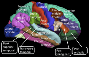
Although there is not an overwhelming amount of research surrounding how the union between craniopagus twins leads to different personality, cognitive and motor traits, there have been some studies exploring what it actually means to share a brain. In the case of Tatiana and Krista as mentioned above, it is possible that the twins shared some conscious thought. Studies of the thalamus’ role in the brain provide neurological data that help explain these behavioral observations that these two twins experience. Thalamo-cortico-thalamic circuits are the looped neural pathways that connect the thalamus to the cerebral cortex, and then the cerebral cortex back to the thalamus. Because the thalamus is mainly responsible for relaying sensory messages from the body to the brain, it is possible that there is a lot of overlap between the twins’ sensory reception and the actual response it creates within the brain. One study examines this by studying the thalamus when it is at a persistent vegetative state that is when the patient is awake but not conscious. This study proved that the cortical activity on its own is not conscious and that all the activity between the loops of the thalamus, the cerebral cortex and the thalamus itself are all conscious actions. Another study of the thalamus reaffirms that the thalamus does not answer yes/no questions but instead acts as a mediator between different parts of the cerebral cortex and systemic sensory reception.[17] These loops actually may account for the relationship between Tatiana and Krista. At the neuronal level, communication is dense network of neurons linked between themselves and the coordinator (in this case the thalamus) that finally sends a message to the cortex. On top of this, there are links between the cortex that send messages back through the coordinator and finally to the rest of the body. The brain’s ability to function through loops and circuits is a good model to explain why Tatiana “consciously” feels what Krista is “physically” experiencing. Additionally there is some level of synchronization between the two twins. Another study found that for craniopagus twins, their connection to each other is comparable to our normal appendages and that their bodies have obvious overlapping physically and psychologically.[18] Because most cases of craniopagus twins are unique, the research outlining general connections between craniopagus twins is limited. However, this example provides insight into the effects of a union between twins who essentially share the same sensory relay system in the thalamus.
History
Conjoined twinning is one of the oldest known birth defects in history and examples with human’s fascination with twins is extremely evident throughout historical literature. The Gemini constellation, known in Greek mythology as Castor and Pollux, is arguably one of the best known sets of twins of all time.[19] In history, Castor and Pollux fought Greek battles alongside other famous war heroes like Herkules and Achilles. The Greeks held these twins in high standing and they were seen not just as warriors but as gods. Although there are cases of conjoined twins dating back to as early as the 10th century, it was not until 1491 that the first case was documented.
Apart from that, Sebastian Munster’s Cosmographia universalis provides the true first account of craniopagus twins who happened to live for ten years, exceeding many expectations during that time.[20] He describes the set of twins as being a unique malformation and a punishment from their mother’s mistake. Furthermore, in Ambroise Pare’s book, On Monsters and Marvels, various types of "supernatural" twinning are illustrated and described as "monstrous and marvelous creatures that proceed from the judgment of God". This published history suggests that conjoined twins, and in specific craniopagus twins, were viewed as literal monsters during that era.
See also
References
- Toreador, A.B., K.L. Cohen, V. Spilotro, and E. Landau. "Craniopagus Twins". Journal of Neurology, Neurosurgery, and Psychiatry (1974): 37. Web
- Browd, Samuel L., James T. Goodrich, and Marion L. Walker. "Craniopagus Twins." Neurosurgery Pediatrics 1.20 (2008): n. pag. Web
- Stone James, Goodrich James (2006). "The Craniopagus Malformation: Classification and Implications for Surgical Separation". Brain. 129 (Pt 5): 1084–95. doi:10.1093/brain/awl065. PMID 16597654.
- Bucholz Richard, Yoon Kong-Woo, Shively Raymond (1987). "Temporparietal Craniopagus". Journal of Neurosurgery. 66 (1): 72–79. doi:10.3171/jns.1987.66.1.0072. PMID 3783261.CS1 maint: multiple names: authors list (link)
- O'Connell, J E (1976). "Craniopagus twins: Surgical anatomy and embryology and their implications". Journal of Neurology, Neurosurgery & Psychiatry. 39: 1–22. doi:10.1136/jnnp.39.1.1. PMC 492208.
- Goodrich, James, and David Staffenberg. "Craniopagus Twins: Clinical and Surgical Management." Child's Nervous System 20.8-9 (2004): 618-24. Web
- Gregory, D. L. (2007). Neurophysiological Studies on Conjoined Twins. In S. A. Smith (Ed.), (Vol. 47): American Journal of Electroneurodiagnostic Technology.]
- Conjoined Twins." Mayo Clinic. N.p., 19 Nov. 2010. Web
- "Finde a Grave record". Retrieved 2017-11-05.
- "Gallery of Famous Craniopagus Twins - Doc Zone - CBC-TV".
- "Egyptian Twins". Retrieved 2008-08-29.
- "Two Lives, Entwined". Archived from the original on 2011-06-07. Retrieved 2012-11-19.
- "NOVA | Separating Twins". Pbs.org. Retrieved 2014-08-03.
- "The battle to separate Safa and Marwa". BBC News. Retrieved 2019-07-17.
- "Beautiful moment conjoined twins see each other for the first time since they were separated". New Zealand Woman's Weekly. November 23, 2016. Retrieved November 27, 2016.
- Dominus, Susan. "Could Conjoined Twins Share a Mind?". The New York Times. Retrieved 21 July 2018.
- Leonard, Abigail. "Your Brain Boots Up Like a Computer." LiveScience (2006): n. pag. Web
- Murray, Craig. "The Experience of Body Boundaries by Siamese Twins." New Ideas in Psychology 19.2 (2001): n. pag. SciVerse. Web
- Squair, Jordan. "Craniopagus: Overview and the Implications of Sharing a Brain." (2012): n. pag. Print.
- Walker, Marion, and Samuel Browd. "Childs Nerve System." Childs Nerve System. 20. (2004): n. page. Web. 19 Nov. 2012.
Further reading
- Stone, James L; Goodrich, James T (2006). "The craniopagus malformation: Classification and implications for surgical separation". Brain. 129 (5): 1084–1095. doi:10.1093/brain/awl065. PMID 16597654.