Fornix (neuroanatomy)
The fornix (meaning "arch" in Latin) is a C-shaped bundle of nerve fibers in the brain that acts as the major output tract of the hippocampus. The fornix also carries some afferent fibres to the hippocampus from structures in the diencephalon and basal forebrain. The fornix is part of the limbic system. While its exact function and importance in the physiology of the brain are still not entirely clear, it has been demonstrated in humans that surgical transection – the cutting of the fornix along its body – can cause memory loss. There is some debate over what type of memory is affected by this damage, but it has been found to most closely correlate with recall memory rather than recognition memory. This means that damage to the fornix can cause difficulty in recalling long-term information such as details of past events, but it has little effect on the ability to recognize objects or familiar situations.
| Fornix | |
|---|---|
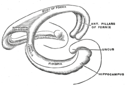 Diagram of the fornix. Right=anterior | |
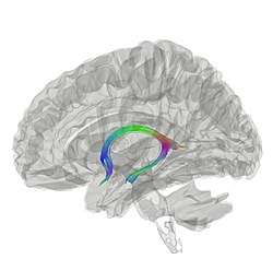 Tractography showing fornix | |
| Details | |
| Artery | Small medial central branches of the anterior communicating artery |
| Identifiers | |
| Latin | Fornix |
| MeSH | D020712 |
| NeuroNames | 268 |
| NeuroLex ID | birnlex_705 |
| TA | A14.1.08.949 A14.1.09.255 |
| FMA | 61965 |
| Anatomical terms of neuroanatomy | |
Structure
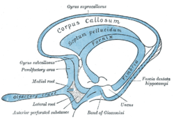
The fibers begin in the hippocampus on each side of the brain as fimbriae; the separate left and right sides are each called the crus of the fornix (plural crura). The bundles of fibers come together in the midline of the brain, forming the body of the fornix. The lower edge of the septum pellucidum (the membrane that separates the lateral ventricles) is attached to the upper face of the fornix body.
The body of the fornix travels anteriorly and divides again near the anterior commissure. The left and right parts separate, but there is also an anterior/posterior divergence.
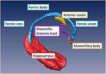
- The posterior fibers (called the postcommissural fornix) of each side continue through the hypothalamus to the mammillary bodies; then to the anterior nuclei of thalamus, which project to the cingulate cortex.
- The anterior fibers (precommissural fornix) end at the septal nuclei and nucleus accumbens of each half of the brain.
Commissure
The lateral portions of the body of the fornix are joined by a thin triangular lamina, named the psalterium (lyra). This lamina contains some commissural fibers that connect the two hippocampi across the middle line and constitute the commissure of fornix (also called the hippocampal commissure).
The terminal lamina creates the commissure plate. This structure gives existence to the corpus callosum, the septum pellucidum, and the fornix. The fornix splits into two columns at the front (anterior pillars), and then splits into two posterior crura. These two crura are joined together through the hippocampal commissure. The beginning of the splitting is called the psalterium or Lyra Davidis. The latter name is used because the structure resembles a lyra (or triangular harp): The two crura are the "chassis" of the lyra, and the commissure connections are the fibers.
Columns
The columns (anterior pillars; fornicolumns) of the fornix arch downward in front of the interventricular foramina and behind the anterior commissure, and each descends through the grey matter in the lateral wall of the third ventricle to the base of the brain, where it ends in the mammillary bodies.
Crus
The crura (posterior pillars) of the fornix are prolonged backward from the body.
They are flattened bands, and, at their commencement, are intimately connected with the under surface of the corpus callosum.
Diverging from one another, each curves around the posterior end of the thalamus, and passes downward and forward into the temporal horn of lateral ventricle.
Here, it lies along the concavity of the hippocampus, on the surface of which some of its fibers are spread out to form the alveus, while the remainder are continued as a narrow white band, the fimbria of hippocampus, which is prolonged into the uncus of the parahippocampal gyrus.
Function
It is known that the fornix is important in some aspects of memory, although there is an ongoing debate as to which in particular. Fornix transection studies in macaques[1] have shown that the monkeys were strongly impaired on object-in-scene learning, which is a type of recall memory, specifically episodic-like memory (integrating what+where, although not when). Similar results were obtained from patients who had had their fornix transected inadvertently during a removal of colloid cysts from their third ventricles.[2] These patients were strongly impaired on general delayed recall tasks, specifically on the Doors and People test, implying an impairment of recall memory, albeit their recognition memory was somewhat impaired as well.
Additional images
 Fornix (animation. click to move)
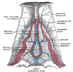 Velum interpositum.
 Fornix
 Fornix
References
This article incorporates text in the public domain from page 837 of the 20th edition of Gray's Anatomy (1918)
- Gaffan, D. (1994). "Scene-specific memory for objects: a model of episodic memory impairment in monkeys with fornix transection". Journal of Cognitive Neuroscience. 6 (4): 305–320. doi:10.1162/jocn.1994.6.4.305. ISSN 0898-929X. PMID 23961727.
- Aggleton, J. P.; McMackin, D.; Carpenter, K.; Hornak, J.; Kapur, N.; Halpin, S.; Wiles, C. M.; Kamel, H.; Brennan, P.; Carton, S.; Gaffan, D. (2000-04-01). "Differential cognitive effects of colloid cysts in the third ventricle that spare or compromise the fornix". Brain. 123 (4): 800–815. doi:10.1093/brain/123.4.800. ISSN 0006-8950. PMID 10734011.
External links
| Wikimedia Commons has media related to Fornix of the brain. |
- More info at BrainInfo
| Authority control |
|---|