Circulating tumor cell
A circulating tumor cell (CTC) is a cell that has shed into the vasculature or lymphatics[1] from a primary tumor and is carried around the body in the blood circulation. CTCs can extravasate and become seeds for the subsequent growth of additional tumors (metastases) in distant organs, a mechanism that is responsible for the vast majority of cancer-related deaths.[2] The detection and analysis of CTCs can assist early patient prognoses and determine appropriate tailored treatments.[3] Currently, there is one FDA-approved method for CTC detection, CellSearch, which is used to diagnose breast, colorectal and prostate cancer.[4]
The detection of CTCs, or liquid biopsy, presents several advantages over traditional tissue biopsies. They are non-invasive, can be used repeatedly, and provide more useful information on metastatic risk, disease progression, and treatment effectiveness.[5][6] For example, analysis of blood samples from cancer patients has found a propensity for increased CTC detection as the disease progresses.[7] Blood tests are easy and safe to perform and multiple samples can be taken over time. By contrast, analysis of solid tumors necessitates invasive procedures that might limit patient compliance. The ability to monitor the disease progression over time could facilitate appropriate modification to a patient's therapy, potentially improving their prognosis and quality of life. The important aspect of the ability to prognose the future progression of the disease is elimination (at least temporarily) of the need for a surgery when the repeated CTC counts are low and not increasing; the obvious benefits of avoiding the surgery include avoiding the risk related to the innate tumor-genicity of cancer surgeries. To this end, technologies with the requisite sensitivity and reproducibility to detect CTCs in patients with metastatic disease have recently been developed.[8][9][10][11][12][13][14][15] On the other hand, CTCs are very rare, often present as only a few cells per milliliter of blood, which makes their detection rather challenging. In addition, they often express a variety of markers which vary from patient to patient, which makes it difficult to develop techniques with high sensitivity and specificity.
Types
CTCs that originate from carcinomas (cancers of epithelial origin, which are the most prevalent) can be classified according to the expression of epithelial markers, as well as their size and whether they are apoptotic. In general, CTCs are anoikis-resistant, which means that they can survive in the bloodstream without attaching to a substrate.[16]
- Traditional CTCs are characterised by an intact, viable nucleus; the expression of EpCAM and cytokeratins, which demonstrate epithelial origin; the absence of CD45, indicating the cell is not of hematopoietic origin; and their larger size, irregular shape or subcellular morphology.[17]
- Cytokeratin-negative CTCs are characterised by the lack of EpCAM or cytokeratins, which may indicate an undifferentiated phenotype (circulating cancer stem cells) or the acquisition of a mesenchymal phenotype (known as epithelial-mesenchymal transition or EMT). These populations of CTCs may be the most resistant and most prone to metastasis. They are also more difficult to isolate because they express neither cytokeratins nor CD45. Otherwise, their morphology, gene expression and genomics are similar to those of other cancer cells.[18]
- Apoptotic CTCs are traditional CTCs that are undergoing apoptosis (programmed cell death). These may be used to monitor treatment response, as done experimentally by the Epic Sciences method, which identifies nuclear fragmentation or cytoplasmic blebbing associated with apoptosis. Measuring the ratio of traditional CTC to apoptotic CTCs—from baseline to therapy—provides clues to treatment efficacy in targeting and killing cancer cells.[18]
- Small CTCs are cytokeratin-positive and CD45-negative, but with sizes and shapes similar to white blood cells. Importantly, small CTCs have cancer-specific biomarkers that identify them as CTCs. Small CTCs have been implicated in progressive disease and differentiation into small cell carcinomas, which often require a different therapeutic course.[19]
CTC clusters
CTC clusters are two or more individual CTCs bound together. The CTC cluster may contain traditional, small or CK- CTCs. These clusters have cancer-specific biomarkers that identify them as CTCs. Several studies have reported that the presence of these clusters is associated with increased metastatic risk and poor prognosis. For example, one study involving prostate cancer showed an eight-fold longer mean survival rate for patients with only single CTCs versus those with CTC clusters, while other studies have shown similar correlations for colon cancer.[20][21] In addition, enumerating CTC clusters can provide useful prognostic information for patients with already elevated CTC levels.[22]
However, one study has reported that contrary to existing consensus, at least a discrete population of these clusters are non-malignant, and derive instead from the tumor endothelium.[23] These circulating tumor-endothelial clusters also show epithelial-mesenchymal markers but do not mirror the genetics of the primary tumor.
Previously it was assumed that CTC clusters could not pass through narrow vessels, such as capillaries, due to their overall size. However, it has been shown that CTC clusters can "unwind" through "selective cleavage of intercellular adhesions" to traverse these constrictions single-file, then reverse the process once clear. This behavior could be a factor in why CTC clusters have such a significant metastatic potential.[24]
Frequency
The detection of CTCs may have important prognostic and therapeutic implications but because their numbers can be very small, these cells are not easily detected.[25] It is estimated that among the cells that have detached from the primary tumor, only 0.01% can form metastases.[26]
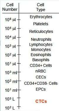
Circulating tumor cells are found in frequencies on the order of 1-10 CTC per mL of whole blood in patients with metastatic disease.[27] For comparison, a mL of blood contains a few million white blood cells and a billion red blood cells. This low frequency, associated to difficulty of identifying cancerous cells, means that a key component of understanding CTCs biological properties require technologies and approaches capable of isolating 1 CTC per mL of blood, either by enrichment, or better yet with enrichment-free assays that identify all CTC subtypes in sufficiently high definition to satisfy diagnostic pathology image-quantity requirements in patients with a variety of cancer types.[18] To date CTCs have been detected in several epithelial cancers (breast, prostate, lung, and colon)[28][29][30][31] and clinical evidences indicate that patients with metastatic lesions are more likely to have CTCs isolated.
CTCs are usually (in 2011) captured from the vasculature by using specific antibodies able to recognize specific tumoral marker (usually EpCAM); however this approach is biased by the need for a sufficient expression of the selected protein on the cell surface, event necessary for the enrichment step. Moreover, since EpCAM and other proteins (e.g. cytokeratins) are not expressed in some tumors and can be down regulated during the epithelial to mesenchymal transition (EMT), new enrichment strategies are required.[32]
First evidence indicates that CTC markers applied in human medicine are conserved in other species. Five of the more common markers including CK19 are also useful to detect CTC in the blood of dogs with malignant mammary tumors.[33][34] Newer approaches are able to identify more cells out 7.5 ml of blood, like IsofFux or Maintrac.[35][36] In very rare cases, CTCs are present in large enough quantities to be visible on routine blood smear examination. This is referred to as carcinocythemia or carcinoma cell leukemia and is associated with a poor prognosis.[37]
Detection methods
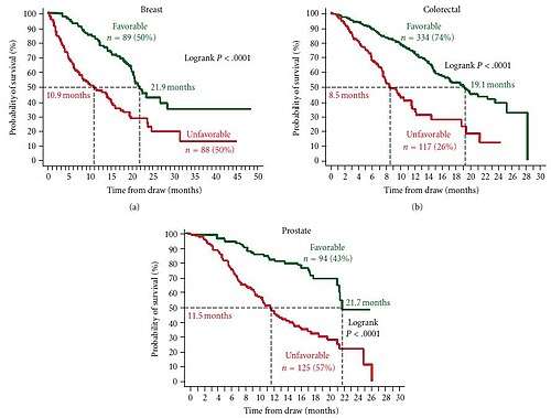
To date, a variety of research methods have been developed to isolate and enumerate CTCs.[38] The only U.S. Food and Drug Administration (FDA) cleared methodology for enumeration of CTC in whole blood is the CellSearch system.[39] Extensive clinical testing done using this method shows that presence of CTCs is a strong prognostic factor for overall survival in patients with metastatic breast, colorectal or prostate cancer.[7][40][41][42][43][44][45]
CTCs are pivotal to understanding the biology of metastasis and promise potential as a biomarker to noninvasively evaluate tumor progression and response to treatment. However, isolation and characterization of CTCs represent a major technological challenge, since CTCs make up a minute number of the total cells in circulating blood, 1–10 CTCs per mL of whole blood compared to a few million white blood cells and a billion red blood cells.[46] Therefore, the major challenge for CTC researchers is the prevailing difficulty of CTC purification that allows the molecular characterization of CTCs. Several methods have been developed to isolate CTCs in the peripheral blood and essentially fall into two categories: biological methods and physical methods, as well as hybrid methods that combine both strategies. Techniques may also be classified based on whether they select CTCs for isolation (positive selection) or whether they exclude all blood cells (negative selection).
Biological methods
Biological methods isolate cells based on highly specific antigen binding, most commonly by monoclonal antibodies for positive selection. Antibodies against tumor specific biomarkers including EpCAM, HER2 and PSA have been used. The most common technique is magnetic nanoparticle-based separation (immunomagnetic assay) as used in CellSearch or MACS. Other techniques under research include microfluidic separation[47] and combination of immunomagnetic assay and microfluidic separation.[48][49][50][51] As the development of microfabrication technology, microscale magnetic structures are implemented to provide better control of the magnetic field and assist the CTCs detection.[52][53][54] Oncolytic viruses such as vaccinia viruses[55] are developed to detect and identify CTCs. Alternative methods exist which use engineered proteins instead of antibodies, such as the malaria VAR2CSA protein, which binds to oncofetal chondroitin sulfate on the surface of CTCs.[56] CTCs may also be retrieved directly from the blood by a modified Seldinger technique, as developed by GILUPI GmbH.[57][58] An antibody coated metal wire is inserted into a peripheral vein and stays there for a defined period (30 min). During this time, CTCs from the blood can bind to the antibodies (currently anti-EpCAM). After the incubation time, the wire is removed, washed and the native CTCs, isolated from the blood of the patient, can be further analysed. Molecular genetics as well as immunofluorescent staining and several other methods are possible.[59][60] Advantage of this method is the higher blood volume that can be analysed for CTCs (approx. 750 ml in 30 min compared to 7.5 ml of a drawn blood sample).
CellSearch method
CellSearch is the only FDA-approved platform for CTC isolation. This method is based on the use of iron nanoparticles coated with a polymer layer carrying biotin analogues and conjugated with antibodies against EpCAM for the capture of CTCs. Isolation is coupled to an analyzer to take images of isolated cells upon their staining with specific fluorescent antibody conjugates. Blood is sampled in an EDTA tube with an added preservative. Upon arrival in the lab, 7.5mL of blood is centrifuged and placed in a preparation system. This system first enriches the tumor cells immunomagnetically by means of ferrofluid nanoparticles and a magnet. Subsequently, recovered cells are permeabilized and stained with a nuclear stain, a fluorescent antibody conjugate against CD45 (leukocyte marker) and cytokeratins 8, 18 and 19 (epithelial markers). The sample is then scanned on an analyzer which takes images of the nuclear, cytokeratin, and CD45 stains.[61] To be considered a CTC a cell must contain a nucleus, be positive for cytoplasmic expression of cytokeratin as well as negative for the expression of CD45 marker, and have a diameter larger than 5 µm. If the total number of tumor cells found to meet the criteria cited above is 5 or more, a blood sample is positive. In studies done on prostate, breast and colon cancer patients, median survival of metastatic patients with positive samples is about half the median survival of metastatic patients with negative samples. This system is characterized by a recovery capacity of 93% and a detection limit of one CTC per 7.5 mL of whole blood. For specific cancer types, alternative methods such as IsoFlux have shown greater sensitivity.[62]
Epic Sciences method
This method involves technology to separate nucleated cells from red blood cells, which lack a nucleus. All nucleated cells, including normal white blood cells and CTCs, are exposed to fluorescent-tagged antibodies specific for cancer biomarkers. In addition, Epic's imaging system captures pictures of all the cells on the slide (approximately 3 million), records the precise coordinates of each cell, and analyzes each cell for 90 different parameters, including the fluorescence intensity of the four fluorescent markers and 86 different morphological parameters. Epic can also use FISH and other staining techniques to look for abnormalities such as duplications, deletions, and rearrangements. The imaging and analysis technology also allows for the coordinates of every cell on a slide to be known so that a single cell can be retrieved from the slide for analysis using next-generation sequencing. A hematopathology-trained algorithm incorporates numerous morphology measurements as well as expression from cytokeratin and CD45. The algorithm then proposes candidate CTCs that a trained reader confirms. Cells of interest are analyzed for relevant phenotypic and genotypic markers, with regional white blood cells included as negative controls.[63] Epic's molecular assays measure protein expression and also interrogate genomic abnormalities in CTCs for more than 20 different cancer types.
Maintrac
Maintrac is a diagnostic blood test platform applying microscopic in vitro diagnostic methods to identify rare cells in body fluids and their molecular characteristics. It is based on positive selection using EpCAM-specific antibodies.[64] Maintrac uses an approach based on microscopic identification of circulating tumor cells. To prevent damage and loss of the cells during the process, Maintrac uses just two steps towards the identification. In contrast to many other methods, maintrac does not purify the cells or enriches them, but identifies them within the context of the other blood compounds. To obtain vital cells and to reduce stress of those cells, blood cells are prepared by only one centrifugation step and erythrocyte lysis. Like CellSearch, maintrac uses an EpCAM antibody. It is, however, not used for enrichment but rather as a fluorescent marker to identify those cells. Together with the nuclear staining with propidium iodide the maintrac method can distinguish between dead and living cells. Only vital, propidium excluding EpCAM positive cells are counted as potential tumor cells. Only living cells can grow into tumors, therefore dying EpCAM positive cells can do no harm. The suspension is analysed by fluorescence microscopy, which automatically counts the events. Simultaneous event galleries are recorded to verify whether the software found a true living cell and to differentiate between skin epithelial cells for example. Close validation of the method showed that additional antibodies of cytokeratins or CD45 did not have any advantage.[36][65]
Unlike other methods maintrac does not use the single cell count as a prognostic marker, rather Maintrac utilizes the dynamics of the cell count. Rising tumor cell numbers are an important factor that tumor activity is ongoing.[66] Decreasing cell counts are a sign for a successful therapy. Therefore, maintrac can be used to verify the success of a chemotherapy[36][67] and to supervise the treatment during hormone or maintenance therapy[68][69] Maintrac has been used experimentally to monito cancer recurrence.[70][71] Studies using Maintrac have shown that EpCAM positive cells can be found in the blood in patient without cancer.[72] Inflammatory conditions like Crohn's disease also show increased levels of EpCAM-positive cells. Patients with severe skin burns can also carry EpCAM positive cells in the blood. Therefore, the use of EpCAM-positive cells as a tool for early diagnosis is not optimal.
Physical methods
Physical methods are often filter-based, enabling the capture of CTCs by size rather than by specific epitopes.[15] ScreenCell is a filtration based device that allows sensitive and specific isolation of CTCs from human whole blood in a few minutes.[73] Peripheral blood is drawn and processed within 4 hours with a ScreenCell isolation device to capture CTCs. The captured cells are ready for cell culture or for direct characterization using ViewRNA in situ hybridization assay. The Parsortix method separates CTCs based on their size and deformability.[74]
Hybrid methods
Hybrid methods combine physical separation (by gradients, magnetic fields, etc.) with antibody-mediated cell retrieval. An example of this is a sensitive double gradient centrifugation and magnetic cell sorting detection and enumeration method which has been used to detect circulating epithelial cancer cells in breast cancer patients by negative selection.[75] The principle of negative selection is based on the retrieval of all blood cells by using a panel of antibodies as well as traditional gradient centrifugation with Ficoll. A similar method known as ISET Test has been employed to detect circulating prostate cancer cells[76][77][78] and another technique known as RosetteStep has been used to isolate CTCs from small-cell lung cancer patients.[79] Similarly, researchers at Massachusetts General Hospital have developed a negative selection method which employs inertial focusing on a microfluidic device. The technique, called CTC-iChip, first removes cells too small to be CTCs, such as red blood cells, and then uses magnetic particles to remove white blood cells.[80]
CTC characterization
Some drugs are particularly effective against cancers which fit certain requirements. For example, Herceptin is very effective in patients who are Her2 positive, but much less effective in patients who are Her2 negative. Once the primary tumor is removed, biopsy of the current state of the cancer through traditional tissue typing is not possible anymore.[81] Often tissue sections of the primary tumor, removed years prior, are used to do the typing. Further characterization of CTC may help determining the current tumor phenotype. FISH assays have been performed on CTC as well as determination of IGF-1R, Her2, Bcl-2, ERG, PTEN, AR status using immunofluorescence.[6][82][83][84][85] Single cell level qPCR can also be performed with the CTCs isolated from blood.
The organ tropism of patient-derived CTC has been investigated in a mouse model.[86] CTCs isolated from breast cancer patients and expanded in vitro showed they could generate bone, lung, ovary and brain metastases in mice, partially reflecting the secondary lesions as found in the corresponding patients. Remarkably, one CTC line—isolated long before the appearance of brain metastasis in patient—was highly competent to generate brain metastasis in mice. This was the first predictive case for brain metastasis and a proof of concept that intrinsic molecular features of metastatic precursors amongst CTCs could provide novel insights into the mechanisms of metastasis.
Cell morphology
Morphological appearance is judged by human operators and is therefore subject to large inter operator variation.[87] Several CTC enumeration methods exist which use morphological appearance to identify CTC, which may also apply different morphological criteria. A recent study in prostate cancer showed that many different morphological definitions of circulating tumor cells have similar prognostic value, even though the absolute number of cells found in patients and normal donors varied by more than a decade between different morphological definitions.[88]
History
CTCs were observed for the first time in 1869 in the blood of a man with metastatic cancer by Thomas Ashworth, who postulated that "cells identical with those of the cancer itself being seen in the blood may tend to throw some light upon the mode of origin of multiple tumours existing in the same person". A thorough comparison of the morphology of the circulating cells to tumor cells from different lesions led Ashworth to conclude that "One thing is certain, that if they [CTC] came from an existing cancer structure, they must have passed through the greater part of the circulatory system to have arrived at the internal saphena vein of the sound leg".[89]
The importance of CTCs in modern cancer research began in the mid 1990s with the demonstration that CTCs exist early on in the course of the disease.[90] Those results were made possible by exquisitely sensitive magnetic separation technology employing ferrofluids (colloidal magnetic nanoparticles) and high gradient magnetic separators invented by Paul Liberti and motivated by theoretical calculations by Liberti and Leon Terstappen that indicated very small tumors shedding cells at less than 1.0% per day should result in detectable cells in blood.[91] A variety of other technologies have been applied to CTC enumeration and identification since that time.
Modern cancer research has demonstrated that CTCs derive from clones in the primary tumor, validating Ashworth's remarks.[92] The significant efforts put into understanding the CTCs biological properties have demonstrated the critical role circulating tumor cells play in the metastatic spread of carcinoma.[93] Furthermore, highly sensitive, single-cell analysis demonstrated a high level of heterogeneity seen at the single cell level for both protein expression and protein localization[94] and the CTCs reflected both the primary biopsy and the changes seen in the metastatic sites.[95]
References
- Riquet, M; Rivera, C; Gibault, L; Pricopi, C; Mordant, P; Badia, A; Arame, A; Le Pimpec Barthes, F (2014). "[Lymphatic spread of lung cancer: anatomical lymph node chains unchained in zones]". Revue de Pneumologie Clinique. 70 (1–2): 16–25. doi:10.1016/j.pneumo.2013.07.001. PMID 24566031.
- Gupta, GP; Massagué, J (Nov 17, 2006). "Cancer metastasis: building a framework". Cell. 127 (4): 679–95. doi:10.1016/j.cell.2006.11.001. PMID 17110329.
- Rack B, Schindlbeck C, Jückstock J, Andergassen U, Hepp P, Zwingers T, Friedl T, Lorenz R, Tesch H, Fasching P, Fehm T, Schneeweiss A, Lichtenegger W, Beckmann M, Friese K, Pantel K, Janni W (2014). "Circulating Tumor Cells Predict Survival in Early Average-to-High Risk Breast Cancer Patients". Journal of the National Cancer Institute. 106 (5). doi:10.1093/jnci/dju066. PMC 4112925. PMID 24832787.
- Millner, LM; Linder, MW; Valdes R, Jr (NaN). "Circulating tumor cells: a review of present methods and the need to identify heterogeneous phenotypes". Annals of Clinical and Laboratory Science. 43 (3): 295–304. PMC 5060940. PMID 23884225. Check date values in:
|date=(help) - Marrinucci, D; Bethel, K; Luttgen, M; Nieva, J; Kuhn, P; Kuhn, P (Sep 2009). "Circulating tumor cells from well-differentiated lung adenocarcinoma retain cytomorphologic features of primary tumor type". Archives of Pathology & Laboratory Medicine. 133 (9): 1468–71. doi:10.1043/1543-2165-133.9.1468 (inactive 2019-12-01). PMC 4422331. PMID 19722757.
- Attard G, Swennenhuis JF, Olmos D, Reid AH, Vickers E, A'Hern R, Levink R, Coumans F, Moreira J, Riisnaes R, Oommen NB, Hawche G, Jameson C, Thompson E, Sipkema R, Carden CP, Parker C, Dearnaley D, Kaye SB, Cooper CS, Molina A, Cox ME, Terstappen LW, de Bono JS (2009). "Characterization of ERG, AR and PTEN gene status in circulating tumor cells from patients with castration-resistant prostate cancer". Cancer Res. 69 (7): 2912–8. doi:10.1158/0008-5472.CAN-08-3667. PMID 19339269.
- Cohen SJ, Punt CJ, Iannotti N, Saidman BH, Sabbath KD, Gabrail NY, Picus J, Morse M, Mitchell E, Miller MC, Doyle GV, Tissing H, Terstappen LW, Meropol NJ (2008). "Relationship of circulating tumor cells to tumor response, progression-free survival, and overall survival in patients with metastatic colorectal cancer". J. Clin. Oncol. 26 (19): 3213–21. doi:10.1200/JCO.2007.15.8923. PMID 18591556.
- Yu M, Ting DT, Stott SL, Wittner BS, Ozsolak F, Paul S, Ciciliano JC, Smas ME, Winokur D, Gilman AJ, Ulman MJ, Xega K, Contino G, Alagesan B, Brannigan BW, Milos PM, Ryan DP, Sequist LV, Bardeesy N, Ramaswamy S, Toner M, Maheswaran S, Haber DA (2012). "RNA sequencing of pancreatic circulating tumour cells implicates WNT signalling in metastasis". Nature. 487 (7408): 510–3. Bibcode:2012Natur.487..510Y. doi:10.1038/nature11217. PMC 3408856. PMID 22763454.
- Sleijfer S, Gratama JW, Sieuwerts AM, et al. (2007). "Circulating tumour cell detection on its way to routine diagnostic implementation?". Eur J Cancer. 43 (18): 2645–50. doi:10.1016/j.ejca.2007.09.016. PMID 17977713.
- Hayes DF, Smerage J.; Smerage (2008). "Is There a Role for Circulating Tumor Cells in the Management of Breast Cancer?". Clin Cancer Res. 14 (12): 3646–50. doi:10.1158/1078-0432.CCR-07-4481. PMID 18559576.
- Pantel K, Alix-Panabières C, Riethdorf S (2009). "Cancer micrometastases". Nat Rev Clin Oncol. 6 (6): 339–51. doi:10.1038/nrclinonc.2009.44. PMID 19399023.
- Pantel K, Riethdorf S.; Riethdorf (2009). "Pathology: are circulating tumor cells predictive of overall survival?". Nature Reviews Clinical Oncology. 6 (4): 190–1. doi:10.1038/nrclinonc.2009.23. PMID 19333222.
- Panteleakou Z, Lembessis P, Sourla A, et al. (2009). "Detection of circulating tumor cells in prostate cancer patients: methodological pitfalls and clinical relevance". Mol Med. 15 (3–4): 101–14. doi:10.2119/molmed.2008.00116. PMC 2600498. PMID 19081770.
- Esmaeilsabzali H, Beischlag TV, Cox ME, Parameswaran AM, Park EJ (2013). "Detection and isolation of circulating tumor cells: principles and methods". Biotechnol. Adv. 31 (7): 1063–84. doi:10.1016/j.biotechadv.2013.08.016. PMID 23999357.
- Nieva, J; Wendel, M; Luttgen, MS; Marrinucci, D; Bazhenova, L; Kolatkar, A; Santala, R; Whittenberger, B; Burke, J; Torrey, M; Bethel, K; Kuhn, P (Feb 2012). "High-definition imaging of circulating tumor cells and associated cellular events in non-small cell lung cancer patients: a longitudinal analysis". Physical Biology. 9 (1): 016004. Bibcode:2012PhBio...9a6004N. doi:10.1088/1478-3975/9/1/016004. PMC 3388002. PMID 22306961.
- Hong, Yupeng; Fang, Francia; Zhang, Qi (December 2016). "Circulating tumor cell clusters: What we know and what we expect (Review)". International Journal of Oncology. 49 (6): 2206–2216. doi:10.3892/ijo.2016.3747. PMC 5117994. PMID 27779656.
- Racila, E; Euhus, D; Weiss, AJ; Rao, C; McConnell, J; Terstappen, LW; Uhr, JW (Apr 1998). "Detection and characterization of carcinoma cells in the blood". Proceedings of the National Academy of Sciences. 95 (8): 4589–4594. Bibcode:1998PNAS...95.4589R. doi:10.1073/pnas.95.8.4589. PMC 22534. PMID 9539782.
- Marrinucci, Dena; Bethel, Kelly; Kolatkar, Anand; Luttgen, Madelyn; Malchiodi, Michael; Baehring, Franziska; Voigt, Katharina; Lazar, Daniel; Nieva, Jorge; Bazhenova, Lyudmilda; Ko, Andrew; Korn, W. Michael; Schram, Ethan; Coward, Michael; Yang, Xing; Metzner, Thomas; Lamy, Rachelle; Honnatti, Meghana; Yoshioka, Craig; Kunken, Joshua; Petrova, Yelena; Sok, Devin; Nelson, David; Kuhn, Peter (Feb 2012). "Fluid Biopsy in Patients with Metastatic Prostate, Pancreatic and Breast Cancers". Physical Biology. 9 (1): 016003. Bibcode:2012PhBio...9a6003M. doi:10.1088/1478-3975/9/1/016003. PMC 3387996. PMID 22306768.
- Ferraldeschi, Roberta; McDaniel, Andrew; Krupa, Rachel; Louw, Jessica; Tucker, Eric; Bales, Natalee; Marrinucci, Dena; Riisnaes, Ruth; Mateo, Joaquin; Dittamore, Ryan; De Bono, Johann Sebastian; Tomlins, Scott A.; Attard, Gerhardt (February 2014). "CK- and small nuclear size circulating tumor cell (CTCs) phenotypes in metastatic castration-resistant prostate cancer (mCRPC)". Journal of Clinical Oncology. 32 (4_suppl): 209. doi:10.1200/jco.2014.32.4_suppl.209.
- Aceto N, Bardia A, Miyamoto DT, Donaldson MC, Wittner BS, Spencer JA, Yu M, Pely A, Engstrom A, Zhu H, Brannigan BW, Kapur R, Stott SL, Shioda T, Ramaswamy S, Ting DT, Lin CP, Toner M, Haber DA, Maheswaran S (2014). "Circulating tumor cell clusters are oligoclonal precursors of breast cancer metastasis". Cell. 158 (5): 1110–22. doi:10.1016/j.cell.2014.07.013. PMC 4149753. PMID 25171411.
- Divella R, Daniele A, Abbate I, Bellizzi A, Savino E, Simone G, Giannone G, Giuliani F, Fazio V, Gadaleta-Caldarola G, Gadaleta C, Lolli I, Sabbà C, Mazzocca A (2014). "The presence of clustered circulating tumor cells (CTCs) and circulating cytokines define an aggressive phenotype in metastatic colorectal cancer". Cancer Causes Control. 25 (11): 1531–41. doi:10.1007/s10552-014-0457-4. PMID 25135616.
- Ye Z, Mu Z, Wang C, Palazzo JP, Biederman L, Li B, Jaslow R, Avery T, Austin L, Yang H, Cristofanilli M (2016). "Prognostic values of circulating tumor cell (CTC) enumeration and their clusters in advanced breast cancer". Cancer Research. 76 (4 Supplement): P2–08–09. doi:10.1158/1538-7445.SABCS15-P2-08-09.
- Cima, I.; Kong, S. L.; Sengupta, D.; Tan, I. B.; Phyo, W. M.; Lee, D.; Hu, M.; Iliescu, C.; Alexander, I.; Goh, W. L.; Rahmani, M.; Suhaimi, N.-A. M.; Vo, J. H.; Tai, J. A.; Tan, J. H.; Chua, C.; Ten, R.; Lim, W. J.; Chew, M. H.; Hauser, C. A. E.; van Dam, R. M.; Lim, W.-Y.; Prabhakar, S.; Lim, B.; Koh, P. K.; Robson, P.; Ying, J. Y.; Hillmer, A. M.; Tan, M.-H. (2016). "Tumor-derived circulating endothelial cell clusters in colorectal cancer". Science Translational Medicine. 8 (345): 345ra89. doi:10.1126/scitranslmed.aad7369. hdl:10754/615874. ISSN 1946-6234. PMID 27358499.
- Au S, Storey B, Moore J, Tang Q, Chen Y, Javaid S, Sarioglu A, Sullivan R, Madden M, O'Keefe R, Haber D, Maheswaran S, Langenau D, Stott S, Toner M (2016). "Clusters of circulating tumor cells traverse capillary-sized vessels". Proceedings of the National Academy of Sciences. 113 (18): 4937–52. Bibcode:2016PNAS..113.4947A. doi:10.1073/pnas.1524448113. PMC 4983862. PMID 27091969.
- Ghossein RA, Bhattacharya S, Rosai J (1999). "Molecular detection of micrometastases and circulating tumor cells in solid tumors". Clin. Cancer Res. 5 (8): 1950–60. PMID 10473071.
- Zhe, X; Cher M.L.; Bonfil R.D. (2011). "Circulating tumor cells: finding the needle in the haystack". Am J Cancer Res. 1 (6): 740–751. PMC 3195935. PMID 22016824.
- Miller MC, Doyle GV, Terstappen LW (2010). "Significance of Circulating Tumor Cells Detected by the CellSearch System in Patients with Metastatic Breast Colorectal and Prostate Cancer". J Oncol. 2010: 1–8. doi:10.1155/2010/617421. PMC 2793426. PMID 20016752.
- Swaby, RF; Cristofanilli, M (Apr 21, 2011). "Circulating tumor cells in breast cancer: a tool whose time has come of age". BMC Medicine. 9: 43. doi:10.1186/1741-7015-9-43. PMC 3107794. PMID 21510857.
- Danila, DC; Fleisher, M; Scher, HI (Jun 15, 2011). "Circulating tumor cells as biomarkers in prostate cancer". Clinical Cancer Research. 17 (12): 3903–12. doi:10.1158/1078-0432.CCR-10-2650. PMC 3743247. PMID 21680546.
- Tanaka F, Yoneda K, Kondo N, Hashimoto M, Takuwa T, Matsumoto S, Okumura Y, Rahman S, Tsubota N, Tsujimura T, Kuribayashi K, Fukuoka K, Nakano T, Hasegawa S (2009). "Circulating tumor cell as a diagnostic marker in primary lung cancer". Clin. Cancer Res. 15 (22): 6980–6. doi:10.1158/1078-0432.CCR-09-1095. PMID 19887487.
- Negin, BP; Cohen, SJ (Jun 2010). "Circulating tumor cells in colorectal cancer: past, present, and future challenges". Current Treatment Options in Oncology. 11 (1–2): 1–13. doi:10.1007/s11864-010-0115-3. PMID 20143276.
- Mikolajczyk, SD; Millar, LS; Tsinberg, P; Coutts, SM; Zomorrodi, M; Pham, T; Bischoff, FZ; Pircher, TJ (2011). "Detection of EpCAM-Negative and Cytokeratin-Negative Circulating Tumor Cells in Peripheral Blood". Journal of Oncology. 2011: 1–10. doi:10.1155/2011/252361. PMC 3090615. PMID 21577258.
- da Costa A, Oliveira JT, Gärtner F, Kohn B, Gruber AD, Klopfleisch R (2011). "Potential markers for detection of circulating canine mammary tumor cells in the peripheral blood". Vet. J. 190 (1): 165–8. doi:10.1016/j.tvjl.2010.09.027. PMID 21051248.
- da Costa, A (2013). "Multiple RT-PCR markers for the detection of circulating tumour cells of metastatic canine mammary tumours". Veterinary Journal. 196 (1): 34–39. doi:10.1016/j.tvjl.2012.08.021. PMID 23036177.
- Harb, W.; Fan, A.; Tran, T.; Danila, D.C.; Keys, D.; Schwartz, M.; and Ionescu-Zanetti, C. (2013). "Mutational Analysis of Circulating Tumor Cells Using a Novel Microfluidic Collection Device and qPCR Assay". Transl. Oncol. 6 (5): 528–538. doi:10.1593/tlo.13367. PMC 3799195. PMID 24151533.
- Pachmann K.; Camara O.; Kavallaris A.; Krauspe S.; Malarski N.; Gajda M.; Kroll T.; Jorke C.; Hammer U.; Altendorf-Hofmann A.; et al. (2008). "Monitoring the Response of Circulating Epithelial Tumor Cells to Adjuvant Chemotherapy in Breast Cancer Allows Detection of Patients at Risk of Early Relapse". J. Clin. Oncol. 26 (8): 1208–1215. doi:10.1200/JCO.2007.13.6523. PMID 18323545.
- Ronen, Shira; Kroft, Steven H.; Olteanu, Horatiu; Hosking, Paul R.; Harrington, Alexandra M. (2019). "Carcinocythemia: A rare entity becoming more common? A 3-year, single institution series of seven cases and literature review". International Journal of Laboratory Hematology. 41 (1): 69–79. doi:10.1111/ijlh.12924. ISSN 1751-5521. PMID 30216684.
- Paterlini-Brechot P, Benali NL.; Benali (2007). "Circulating tumor cells (CTC) detection: Clinical impact and future directions". Cancer Lett. 253 (2): 180–204. doi:10.1016/j.canlet.2006.12.014. PMID 17314005.
- "Veridex CellSearch Website". March 2010. Archived from the original on 2008-06-05. Retrieved 2010-03-14.
- "Veridex LLC. CellSearch circulating tumor cell kit premarket notification—expanded indications for use—metastatic prostate cancer" (PDF). March 2010. Retrieved 2010-03-14.
- Cristofanilli M, Budd GT, Ellis MJ, et al. (2004). "Circulating Tumor Cells, Disease Progression and Survival in Metastatic Breast Cancer". NEJM. 351 (8): 781–91. doi:10.1056/NEJMoa040766. PMID 15317891.
- Budd G, Cristofanilli M, Ellis M, et al. (2006). "Circulating Tumor Cells versus Imaging - Predicting Overall Survival in Metastatic Breast Cancer". Clin Can Res. 12 (21): 6404–09. doi:10.1158/1078-0432.CCR-05-1769. PMID 17085652.
- JS DeBono; HI Scher; RB Montgomery; et al. (2008). "Circulating Tumor Cells (CTC) predict survival benefit from treatment in metastatic castration resistant prostate cancer (CRPC)". Clin Can Res. 14 (19): 6302–9. doi:10.1158/1078-0432.CCR-08-0872. PMID 18829513.
- Allard WJ, Matera J, Miller MC, et al. (2004). "Tumor cells circulate in the peripheral blood of all major carcinomas but not in healthy subjects or patients with non-malignant diseases". Clin Can Res. 10 (20): 6897–6904. doi:10.1158/1078-0432.CCR-04-0378. PMID 15501967.
- Riethdorf; Fritsche, H; Müller, V; Rau, T; Schindlbeck, C; Rack, B; Janni, W; Coith, C; et al. (2007). "Detection of Circulating Tumor Cells in Peripheral Blood of Patients with Metastatic Breast Cancer: A Validation Study of the CellSearch System". Clin Cancer Res. 13 (3): 920–8. doi:10.1158/1078-0432.CCR-06-1695. PMID 17289886.
- Yu M.; et al. (2011). "Circulating tumor cells: approaches to isolation and characterization". The Journal of Cell Biology. 192 (3): 373–382. doi:10.1083/jcb.201010021. PMC 3101098. PMID 21300848.
- Nagrath, Sunitha; Sequist, Lecia V.; Maheswaran, Shyamala; Bell, Daphne W.; Irimia, Daniel; Ulkus, Lindsey; Smith, Matthew R.; Kwak, Eunice L.; Digumarthy, Subba; Muzikansky, Alona; Ryan, Paula; Balis, Ulysses J.; Tompkins, Ronald G.; Haber, Daniel A.; Toner, Mehmet (December 2007). "Isolation of rare circulating tumour cells in cancer patients by microchip technology". Nature. 450 (7173): 1235–1239. Bibcode:2007Natur.450.1235N. doi:10.1038/nature06385. PMC 3090667. PMID 18097410.
- Hoshino, Kazunori; Huang, Yu-Yen; Lane, Nancy; Huebschman, Michael; Uhr, Jonathan W.; Frenkel, Eugene P.; Zhang, Xiaojing (Oct 2011). "Microchip-based immunomagnetic detection of circulating tumor cells". Lab on a Chip. 11 (20): 3449–3457. doi:10.1039/c1lc20270g. PMC 3379551. PMID 21863182.
- Peng, Chen; Yu-yen, Huang; Hoshino, Kazunori; Xiaojing, Zhang (2014). "Multiscale immunomagnetic enrichment of circulating tumor cells: from tubes to microchips". Lab on a Chip. 14 (3): 446–458. doi:10.1039/C3LC51107C. PMID 24292816.
- Huang, Yu-yen; Hoshino, Kazunori; Chen, Peng; Wu, Chun-hsien; Lane, Nancy; Huebschman, Michael; Liu, Huaying; Sokolov, Konstantin; Uhr, Jonathan W. (2012-10-30). "Immunomagnetic nanoscreening of circulating tumor cells with a motion controlled microfluidic system". Biomedical Microdevices. 15 (4): 673–681. doi:10.1007/s10544-012-9718-8. ISSN 1387-2176. PMC 3584207. PMID 23109037.
- Hoshino, Kazunori; Chen, Peng; Huang, Yu-Yen; Zhang, Xiaojing (2012-05-15). "Computational Analysis of Microfluidic Immunomagnetic Rare Cell Separation from a Particulate Blood Flow". Analytical Chemistry. 84 (10): 4292–4299. doi:10.1021/ac2032386. ISSN 0003-2700. PMC 3359653. PMID 22510236.
- Chen, Peng; Huang, Yu-Yen; Hoshino, Kazunori; Zhang, John X.J. (2015-03-04). "Microscale Magnetic Field Modulation for Enhanced Capture and Distribution of Rare Circulating Tumor Cells". Scientific Reports. 5: 8745. Bibcode:2015NatSR...5E8745C. doi:10.1038/srep08745. ISSN 2045-2322. PMC 4348664. PMID 25735563.
- Huang, Yu-Yen; Chen, Peng; Wu, Chun-Hsien; Hoshino, Kazunori; Sokolov, Konstantin; Lane, Nancy; Liu, Huaying; Huebschman, Michael; Frenkel, Eugene (2015-11-05). "Screening and Molecular Analysis of Single Circulating Tumor Cells Using Micromagnet Array". Scientific Reports. 5: 16047. Bibcode:2015NatSR...516047H. doi:10.1038/srep16047. ISSN 2045-2322. PMC 4633592. PMID 26538094.
- Chen, Peng; Huang, Yu-Yen; Bhave, Gauri; Hoshino, Kazunori; Zhang, Xiaojing (2015-08-20). "Inkjet-Print Micromagnet Array on Glass Slides for Immunomagnetic Enrichment of Circulating Tumor Cells". Annals of Biomedical Engineering. 44 (5): 1710–1720. doi:10.1007/s10439-015-1427-z. ISSN 0090-6964. PMC 4761332. PMID 26289942.
- Wang, Huiqiang; Chen, Nanhai G.; Minev, Boris R.; Zimmermann, Martina; Aguilar, Richard J.; Zhang, Qian; Sturm, Julia B.; Fend, Falko; Yu, Yong A.; Cappello, Joseph; Lauer, Ulrich M.; Szalay, Aladar A. (September 2013). "Optical Detection and Virotherapy of Live Metastatic Tumor Cells in Body Fluids with Vaccinia Strains". PLoS ONE. 8 (9): e71105. Bibcode:2013PLoSO...871105W. doi:10.1371/journal.pone.0071105. PMC 3760980. PMID 24019862.
- Agerbæk, Mette Ø.; Bang-Christensen, Sara R.; Yang, Ming-Hsin; Clausen, Thomas M.; Pereira, Marina A.; Sharma, Shreya; Ditlev, Sisse B.; Nielsen, Morten A.; Choudhary, Swati; Gustavsson, Tobias; Sorensen, Poul H.; Meyer, Tim; Propper, David; Shamash, Jonathan; Theander, Thor G.; Aicher, Alexandra; Daugaard, Mads; Heeschen, Christopher; Salanti, Ali (16 August 2018). "The VAR2CSA malaria protein efficiently retrieves circulating tumor cells in an EpCAM-independent manner". Nature Communications. 9 (1): 3279. doi:10.1038/s41467-018-05793-2. PMC 6095877. PMID 30115931.
- GILUPI. "GILUPI - CellCollector in vivo CTC isolation". www.gilupi.de.
- Saucedo-Zeni Nadia; et al. (2012). "A novel method for the in vivo isolation of circulating tumor cells from peripheral blood of cancer patients using a functionalized and structured medical wire". International Journal of Oncology. 41 (4): 1241–1250. doi:10.3892/ijo.2012.1557. PMC 3583719. PMID 22825490.
- Luecke, Klaus, et al. "The GILUPI CellCollector as an in vivo tool for circulating tumor cell enumeration and molecular characterization in lung cancer patients." ASCO Annual Meeting Proceedings. Vol. 33. No. 15_suppl. 2015. http://hwmaint.meeting.ascopubs.org/cgi/content/abstract/33/15_suppl/e22035 Archived 2016-03-10 at the Wayback Machine
- Scheumann N; et al. (2015). "50P * Enumeration and Molecular Characterization of Circulating Tumor Cells in Lung Cancer Patients Using the Gilupi Cellcollector , an Effective in Vivo Device for Capturing CTCS". Annals of Oncology. 26: i14. doi:10.1093/annonc/mdv045.14.
- "An Introduction to the CellSearch™" (PDF).
- Sánchez-Lorencio, M.I.; Ramirez, P.; Saenz, L.; Martínez Sánchez, M.V.; De La Orden, V.; Mediero-Valeros, B.; Veganzones-De-Castro, S.; Baroja-Mazo, A.; Revilla Nuin, B.; Gonzalez, M.R.; Cascales-Campos, P.A.; Noguera-Velasco, J.A.; Minguela, A.; Díaz-Rubio, E.; Pons, J.A.; Parrilla, P. (November 2015). "Comparison of Two Types of Liquid Biopsies in Patients With Hepatocellular Carcinoma Awaiting Orthotopic Liver Transplantation". Transplantation Proceedings. 47 (9): 2639–2642. doi:10.1016/j.transproceed.2015.10.003. PMID 26680058.
- Bethel, Kelly; Luttgen2, Madelyn; Damani, Samir; Kolatkar2, Anand; Lamy, Rachelle; Sabouri-Ghomi, Mohsen; Topol, Sarah; Topol2, Eric; Kuhn, Peter (9 Jan 2014). "Fluid phase biopsy for detection and characterization of circulating endothelial cells in myocardial infarction". Physical Biology. 11 (1): 016002. Bibcode:2014PhBio..11a6002B. doi:10.1088/1478-3975/11/1/016002. PMC 4143170. PMID 24406475.
- Pachmann, Katharina (5 April 2015). "Current and potential use of MAINTRAC method for cancer diagnosis and prediction of metastasis". Expert Review of Molecular Diagnostics. 15 (5): 597–605. doi:10.1586/14737159.2015.1032260. PMID 25843106.
- Pachmann K.; Camara O.; Kavallaris A.; Schneider U.; Schünemann S.; Höffken K. (2005). "Quantification of the response of circulating epithelial cells to neodadjuvant treatment for breast cancer: a new tool for therapy monitoring". Breast Cancer Res. 7 (6): R975–979. doi:10.1186/bcr1328. PMC 1410761. PMID 16280045.
- Lobodasch, Kurt; Fröhlich, Frank; Rengsberger, Matthias; Schubert, Rene; Dengler, Robert; Pachmann, Ulrich; Pachmann, Katharina (April 2007). "Quantification of circulating tumour cells for the monitoring of adjuvant therapy in breast cancer: An increase in cell number at completion of therapy is a predictor of early relapse". The Breast. 16 (2): 211–218. doi:10.1016/j.breast.2006.12.005. ISSN 0960-9776. PMID 17291754.
- Camara O.; Rengsberger M.; Egbe A.; Koch A.; Gajda M.; Hammer U.; Jorke C.; Rabenstein C.; Untch M.; Pachmann K. (2007). "The relevance of circulating epithelial tumor cells (CETC) for therapy monitoring during neoadjuvant (primary systemic) chemotherapy in breast cancer". Ann. Oncol. 18 (9): 1484–1492. doi:10.1093/annonc/mdm206. PMID 17761704.
- Pachmann K.; Camara O.; Kohlhase A.; Rabenstein C.; Kroll T.; Runnebaum I.B.; Hoeffken K. (2010). "Assessing the efficacy of targeted therapy using circulating epithelial tumor cells (CETC): the example of SERM therapy monitoring as a unique tool to individualize therapy". J. Cancer Res. Clin. Oncol. 137 (5): 821–828. doi:10.1007/s00432-010-0942-4. PMC 3074080. PMID 20694797.
- Pachmann K.; Camara O.; Kroll T.; Gajda M.; Gellner A.K.; Wotschadlo J.; Runnebaum I.B. (2011). "Efficacy control of therapy using circulating epithelial tumor cells (CETC) as "Liquid Biopsy": trastuzumab in HER2/neu-positive breast carcinoma". J. Cancer Res. Clin. Oncol. 137 (9): 1317–1327. doi:10.1007/s00432-011-1000-6. PMC 3155034. PMID 21739182.
- Hekimian K.; Meisezahl S.; Trompelt K.; Rabenstein C.; Pachmann K. (2012). "Epithelial Cell Dissemination and Readhesion: Analysis of Factors Contributing to Metastasis Formation in Breast Cancer". ISRN Oncol. 2012: 1–8. doi:10.5402/2012/601810. PMC 3317055. PMID 22530147.
- Rolle A.; Günzel R.; Pachmann U.; Willen B.; Höffken K.; Pachmann K. (2005). "Increase in number of circulating disseminated epithelial cells after surgery for non-small cell lung cancer monitored by MAINTRAC is a predictor for relapse: A preliminary report". World J. Surg. Oncol. 3 (1): 18. doi:10.1186/1477-7819-3-18. PMC 1087511. PMID 15801980.
- Camara Oumar; Kavallaris Andreas; Nöschel Helmut; Rengsberger Matthias; Jörke Cornelia; Pachmann Katharina (2006). "Seeding of Epithelial Cells into Circulation During Surgery for Breast Cancer: The Fate of Malignant and Benign Mobilized Cells". World Journal of Surgical Oncology. 4: 67. doi:10.1186/1477-7819-4-67. PMC 1599731. PMID 17002789.
- Desitter I.; et al. (2011). "A New Device for Rapid Isolation by Size and Characterization of Rare Circulating Tumor Cells". Anticancer Research. 31 (2): 427–442.
- Miller, M. Craig; Robinson, Peggy S.; Wagner, Christopher; O'Shannessy, Daniel J. (14 August 2018). "The Parsortix™ Cell Separation System—A versatile liquid biopsy platform". Cytometry Part A. 93 (12): 1234–1239. doi:10.1002/cyto.a.23571. PMID 30107082.
- Tkaczuk KH, Goloubeva O, Tait NS, Feldman F, Tan M, Lum ZP, Lesko SA, Van Echo DA, Ts'o PO (2008). "The significance of circulating epithelial cells in Breast Cancer patients by a novel negative selection method". Breast Cancer Res. Treat. 111 (2): 355–64. doi:10.1007/s10549-007-9771-9. PMID 18064568.
- Wang ZP, Eisenberger MA, Carducci MA, Partin AW, Scher HI, Ts'o PO (2000). "Identification and characterization of circulating prostate carcinoma cells". Cancer. 88 (12): 2787–95. doi:10.1002/1097-0142(20000615)88:12<2787::aid-cncr18>3.0.co;2-2. PMID 10870062.
- Polascik TJ, Wang ZP, Shue M, Di S, Gurganus RT, Hortopan SC, Ts'o PO, Partin AW (1999). "Influence of sextant prostate needle biopsy or surgery on the detection and harvest of intact circulating prostate cancer cells". J. Urol. 162 (3 Pt 1): 749–52. doi:10.1097/00005392-199909010-00034. PMID 10458358.
- Ali A, Furusato B, Ts'o PO, Lum ZP, Elsamanoudi S, Mohamed A, Srivastava S, Moul JW, Brassell SA, Sesterhenn IA, McLeod DG (2010). "Assessment of circulating tumor cells (CTCs) in prostate cancer patients with low-volume tumors". Pathol. Int. 60 (10): 667–72. doi:10.1111/j.1440-1827.2010.02584.x. PMID 20846264.
- Hodgkinson, Cassandra L; Morrow, Christopher J; Li, Yaoyong; Metcalf, Robert L; Rothwell, Dominic G; Trapani, Francesca; Polanski, Radoslaw; Burt, Deborah J; Simpson, Kathryn L; Morris, Karen; Pepper, Stuart D; Nonaka, Daisuke; Greystoke, Alastair; Kelly, Paul; Bola, Becky; Krebs, Matthew G; Antonello, Jenny; Ayub, Mahmood; Faulkner, Suzanne; Priest, Lynsey; Carter, Louise; Tate, Catriona; Miller, Crispin J; Blackhall, Fiona; Brady, Ged; Dive, Caroline (1 June 2014). "Tumorigenicity and genetic profiling of circulating tumor cells in small-cell lung cancer". Nature Medicine. 20 (8): 897–903. doi:10.1038/nm.3600. PMID 24880617.
- Ozkumur E, Shah AM, Ciciliano JC, Emmink BL, Miyamoto DT, Brachtel E, Yu M, Chen PI, Morgan B, Trautwein J, Kimura A, Sengupta S, Stott SL, Karabacak NM, Barber TA, Walsh JR, Smith K, Spuhler PS, Sullivan JP, Lee RJ, Ting DT, Luo X, Shaw AT, Bardia A, Sequist LV, Louis DN, Maheswaran S, Kapur R, Haber DA, Toner M (2013). "Inertial focusing for tumor antigen-dependent and -independent sorting of rare circulating tumor cells". Science Translational Medicine. 5 (179): 179. doi:10.1126/scitranslmed.3005616. PMC 3760275. PMID 23552373.
- Meng S, Tripathy D, Shete S, Ashfaq R, Haley B, Perkins S, Beitsch P, Khan A, Euhus D, Osborne C, Frenkel E, Hoover S, Leitch M, Clifford E, Vitetta E, Morrison L, Herlyn D, Terstappen LW, Fleming T, Fehm T, Tucker T, Lane N, Wang J, Uhr J (2004). "HER-2 gene amplification can be acquired as breast cancer progresses". Proc. Natl. Acad. Sci. U.S.A. 101 (25): 9393–8. Bibcode:2004PNAS..101.9393M. doi:10.1073/pnas.0402993101. PMC 438987. PMID 15194824.
- Hayes DF, Walker TM, Singh B, et al. (2002). "Monitoring Expression of HER-2 on Circulating Epithelial Cells in Patients with advanced Breast Cancer". Int J Oncol. 21: 1111–8.
- O'Hara SM, Moreno JG, Zweitzig DR, et al. (2004). "Multigene Reverse Transcription-PCR Profiling of Circulating Tumor Cells in Hormone-Refractory Prostate Cancer". Clin Chem. 50 (5): 826–835. doi:10.1373/clinchem.2003.028563. PMID 14988224.
- de Bono JS, Attard G, Adjei A, et al. (2007). "Potential Applications for Circulating Tumor Cells expressing the Insulin Growth Factor-I Receptor". Clin Can Res. 13 (12): 3611–6. doi:10.1158/1078-0432.CCR-07-0268. PMID 17575225.
- Karp DD, Pollak MN, Cohen RB, et al. (2009). "Pharmacokinetics and Pharmacodynamics of the IGF-IR Inhibitor Figitumumab (CP-751,871) in Combination with Paclitaxel and Carboplatin". Journal of Thoracic Oncology. 4 (11): 1397–1403. doi:10.1097/JTO.0b013e3181ba2f1d. PMC 2941876. PMID 19745765.
- Klotz, Remi; Thomas, Amal; Teng, Teng; Han, Sung Min; Iriondo, Oihana; Li, Lin; Restrepo-Vassalli, Sara; Wang, Alan; Izadian, Negeen; MacKay, Matthew; Moon, Byoung-San (2019-01-01). "Circulating tumor cells exhibit metastatic tropism and reveal brain metastasis drivers". Cancer Discovery: CD–19–0384. doi:10.1158/2159-8290.CD-19-0384. ISSN 2159-8274. PMID 31601552.
- AGJ Tibbe; MC Miller; LWMM Terstappen (2007). "Statistical Considerations for Enumeration of Circulating Tumor Cells". Cytometry Part A. 71A (3): 132–142. doi:10.1002/cyto.a.20369. PMID 17200956.
- F. A. W. Coumans; C. J. M. Doggen; G. Attard; et al. (2010). "All circulating EpCAM1CK1CD452 objects predict overall survival in castration-resistant prostate cancer". Annals of Oncology. 21 (9): 1851–7. doi:10.1093/annonc/mdq030. PMID 20147742.
- Ashworth, T. R (1869). "A case of cancer in which cells similar to those in the tumours were seen in the blood after death". Australian Medical Journal. 14: 146–7.
- Racila, E.; Euhus, D.; Weiss, A. J.; Rao, C.; McConnell, J.; Terstappen, L. W. M. M.; Uhr, J. W. (1998). "Detection and characterization of carcinoma cells in the blood". Proceedings of the National Academy of Sciences. 95 (8): 4589–4594. Bibcode:1998PNAS...95.4589R. doi:10.1073/pnas.95.8.4589. ISSN 0027-8424. PMC 22534. PMID 9539782.
- Paul Liberti (November 22, 2013). "The tools that fueled the new world of Circulating Tumor Cells by Paul A. Liberti – BioMagnetic Solutions". biomargneticsolutions.com.
- Fehm T, Sagalowsky A, Clifford E, Beitsch P, Saboorian H, Euhus D, Meng S, Morrison L, Tucker T, Lane N, Ghadimi BM, Heselmeyer-Haddad K, Ried T, Rao C, Uhr J (Jul 2002). "Cytogenetic evidence that circulating epithelial cells in patients with carcinoma are malignant". Clinical Cancer Research. 8 (7): 2073–84. PMID 12114406.
- Fidler IJ (2003). "Timeline: The pathogenesis of cancer metastasis: the 'seed and soil' hypothesis revisited". Nature Reviews Cancer. 3 (6): 453–8. doi:10.1038/nrc1098. PMID 12778135.
- "Meeting Library - Meeting Library". meetinglibrary.asco.org.
- Design, ISITE. "OASIS". www.abstractsonline.com.