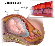
Chorionic villi
Chorionic villi are villi that sprout from the chorion to provide maximal contact area with maternal blood.
| Chorionic villi | |
|---|---|
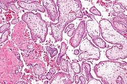 Micrograph showing chorionic villi. Intermediate magnification. H&E stain. | |
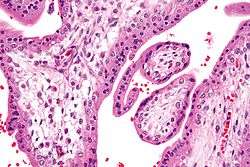 Micrograph showing chorionic villi. Very high magnification. H&E stain. | |
| Details | |
| Days | 24 |
| Identifiers | |
| MeSH | D002824 |
| Anatomical terminology | |
They are an essential element in pregnancy from a histomorphologic perspective, and are, by definition, a product of conception. Branches of the umbilical arteries carry embryonic blood to the villi. After circulating through the capillaries of the villi, blood returns to the embryo through the umbilical vein. Thus, villi are part of the border between maternal and fetal blood during pregnancy.
Structure
Villi can also be classified by their relations:
- Floating villi float freely in the intervillous space. They exhibit a bi-layered epithelium consisting of cytotrophoblasts with overlaying syncytium (syncytiotrophoblast).
- Anchoring (stem) villi stabilize mechanical integrity of the placental-maternal interface.
Development
The chorion undergoes rapid proliferation and forms numerous processes, the chorionic villi, which invade and destroy the uterine decidua and at the same time absorb from it nutritive materials for the growth of the embryo. They undergo several stages, depending on their composition.
| Stage | Description | Period of gestation | Contents |
| Primary | The chorionic villi are at first small and non-vascular. | 13–15 days | trophoblast only[1] |
| Secondary | The villi increase in size and ramify, while the mesoderm grows into them. | 16–21 days | trophoblast and mesoderm[1] |
| Tertiary | Branches of the umbilical artery and umbilical vein grow into the mesoderm, and in this way the chorionic villi are vascularized. | 17-22 days | trophoblast, mesoderm, and blood vessels[1] |
Until about the end of the second month of pregnancy, the villi cover the entire chorion, and are almost uniform in size—but after then, they develop unequally.
Microanatomy
The bulk of the villi consist of connective tissues that contain blood vessels. Most of the cells in the connective tissue core of the villi are fibroblasts. Macrophages known as Hofbauer cells are also present.
Clinical significance
Use for prenatal diagnosis
In 1983, an Italian biologist named Giuseppe Simoni discovered a new method of prenatal diagnosis using chorionic villi.
Stem cell
Chorionic villi are a rich source of stem cells. Biocell Center, a biotech company managed by Giuseppe Simoni, is studying and testing these types of stem cells. Chorionic stem cells, like amniotic stem cells, are uncontroversial multipotent stem cells.[2][3][4]
Infections
Recet studies indicate that the chorionic villi tissues may be susceptible to pathogenic infections,[5] including viral infections. Indeed, footprints of JC polyomavirus and Merkel Cell polyomavirus have been detected in chorionic villi from females affected by spontaneous abortion as well as pregnant women.[6][7] Another virus, BK polyomavirus has been detected in the same tissues, but with lesser extent.[8]
Additional images
 Section through the embryo.
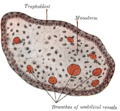 Transverse section of a chorionic villus.
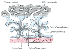 Primary chorionic villi. Diagrammatic.
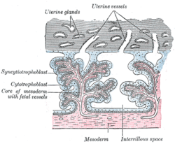 Secondary (Tertiary? Vessels are present.) chorionic villi. Diagrammatic.
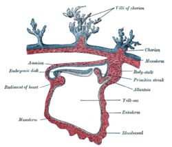
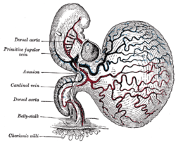 Human embryo of about 28 days, with yolk-sac.
See also
References
This article incorporates text in the public domain from page 60 of the 20th edition of Gray's Anatomy (1918)
- Larsen, William J. : Human embryology. Sherman, Lawrence S.; Potter, S. Steven; Scott, William J. 3. ed.
- "European Biotech Company Biocell Center Opens First U.S. Facility for Preservation of Amniotic Stem Cells in Medford, Massachusetts | Reuters". 2009-10-22. Retrieved 2010-01-11.
- "Europe's Biocell Center opens Medford office - Daily Business Update - The Boston Globe". 2009-10-22. Retrieved 2010-01-11.
- "The Ticker - BostonHerald.com". Retrieved 2010-01-11.
- Contini C, Rotondo JC, Magagnoli F, Maritati M, Seraceni S, Graziano A (2018). "Investigation on silent bacterial infections in specimens from pregnant women affected by spontaneous miscarriage". J Cell Physiol. 34 (3): 433–440. doi:10.1002/jcp.26952. PMID 30078192.
- Tagliapietra A, Rotondo JC, Bononi I, Mazzoni E, Magagnoli F, Maritati M (2019). "Footprints of BK and JC polyomaviruses in specimens from females affected by spontaneous abortion". Hum Reprod. 34 (3): 433–440. doi:10.1002/jcp.27490. PMID 30590693.
- Tagliapietra A, Rotondo JC, Bononi I, Mazzoni E, Magagnoli F, Maritati M (2019). "Droplet-digital PCR assay to detect Merkel cell polyomavirus sequences in chorionic villi from spontaneous abortion affected females". J Cell Physiol. 235 (3): 1888–1894. doi:10.1002/jcp.29213. PMID 31549405.
- Tagliapietra A, Rotondo JC, Bononi I, Mazzoni E, Magagnoli F, Maritati M (2019). "Footprints of BK and JC polyomaviruses in specimens from females affected by spontaneous abortion". Hum Reprod. 34 (3): 433–440. doi:10.1002/jcp.27490. PMID 30590693.