Chordae tendineae
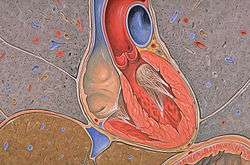
The chordae tendineae (tendinous cords), colloquially known as the heart strings, are tendon-resembling fibrous cords of connective tissue that connect the papillary muscles to the tricuspid valve and the bicuspid valve in the heart.
| Chordae tendineae | |
|---|---|
 The chordae tendineae connect the valves to the heart muscle | |
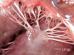 Papillary muscles and chordae tendineae | |
| Details | |
| Identifiers | |
| Latin | chordae tendineae cordis |
| MeSH | D002815 |
| TA | A12.1.00.023 |
| FMA | 76527 |
| Anatomical terminology | |
Chordae tendineae are approximately 80% collagen, while the remaining 20% is made up of elastin and endothelial cells.
Structure
Tendon of Todaro
The tendon of Todaro is a continuation of the Eustachian valve of the inferior vena cava and the Thebesian ring of the coronary sinus. Along with the opening of the coronary sinus and the septal cusp of the tricuspid valve, it makes up the triangle of Koch. The apex of the triangle of Koch is the location of the atrioventricular node.
Function
During atrial systole, blood flows from the atria to the ventricles down the pressure gradient. Chordae tendineae are relaxed because the atrioventricular valves are forced open.[1]
When the ventricles of the heart contract in ventricular systole, the increased blood pressures in both chambers push the AV valves to close simultaneously, preventing backflow of blood into the atria. Since the blood pressure in atria is much lower than that in the ventricles, the flaps attempt to evert to the low pressure regions. The chordae tendineae prevent the eversion, prolapse, by becoming tense thus pulling the flaps, holding them in closed position.[1]
Additional images
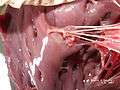 Papillary muscles and chordae tendineae
- Ultrasound showing redundant chordae tendineae[2]
See also
References
- Karas, S.; Elkins, R. C. (1970). "Mechanism of Function of the Mitral Valve Leaflets, Chordae Tendineae and Left Ventricular Papillary Muscles in Dogs". Circulation Research. 26 (6): 689–96. doi:10.1161/01.RES.26.6.689. PMID 5422929.
- "UOTW #75 - Ultrasound of the Week". Ultrasound of the Week. 4 November 2016. Retrieved 27 May 2017.
| Authority control |
|---|