Cerebral angiography
Cerebral angiography is a form of angiography which provides images of blood vessels in and around the brain, thereby allowing detection of abnormalities such as arteriovenous malformations and aneurysms. It was pioneered in 1927 by the Portuguese neurologist Egas Moniz at the University of Lisbon, who also helped develop thorotrast for use in the procedure.[1]
| Cerebral angiography | |
|---|---|
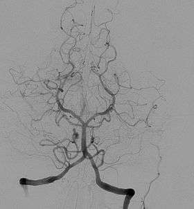 Cerebral angiogram showing a transverse projection of the vertebrobasilar and posterior cerebral circulation. | |
| ICD-9-CM | 88.41 |
| MeSH | D002533 |
| MedlinePlus | 003799 |
Typically a catheter is inserted into a large artery (such as the femoral artery) and threaded through the circulatory system to the carotid artery, where a contrast agent is injected. A series of radiographs are taken as the contrast agent spreads through the brain's arterial system, then a second series as it reaches the venous system.
Prior to the 1970s the typical technique involved a needle puncture directly into the carotid artery,[2][3] as depicted in the 1973 horror film The Exorcist,[4] which was replaced by the current method of threading a catheter from a distant artery due to common complications caused by trauma to the artery at the puncture site in the neck (particularly hematomas of the neck, with possible compromission of the airway).[5][6]
For some applications cerebral angiography may yield better images than less invasive methods such as computed tomography angiography and magnetic resonance angiography. In addition, cerebral angiography allows certain treatments to be performed immediately, based on its findings. In recent decades, cerebral angiography has so assumed a therapeutic connotation thanks to the elaboration of endovascular therapeutic techniques. Embolization (a minimally invasive surgical technique) over time has played an increasingly significant role in the multimodal treatment of cerebral MAVs, facilitating subsequent microsurgical or radiosurgical treatment.[7][8] Another type of treatment possible by angiography (if the images reveal an aneurysm) is the introduction of metal coils through the catheter already in place and maneuvered to the site of aneurysm; over time these coils encourage formation of connective tissue at the site, strengthening the vessel walls.[9][10]
In some jurisdictions, cerebral angiography is required to confirm brain death.
Prior to the advent of modern neuroimaging techniques such as MRI and CT in the mid-1970s, cerebral angiographies were frequently employed as a tool to infer the existence and location of certain kinds of lesions and hematomas by looking for secondary vascular displacement caused by the mass effect related to these medical conditions. This use of angiography as an indirect assessment tool is nowadays obsolete as modern non-invasive diagnostic methods are available to image many kinds of primary intracranial abnormalities directly.[11] It is still widely used however for evaluating various types of vascular pathologies within the skull.
See also

References
- Tondreau, R. (1985) Egas Moniz 1874-1955. Radiographics, 5(6):994-997
- Nii K, Kazekawa K, Onizuka M, Aikawa H, Tsutsumi M, Tomokiyo M, Iko M, Kodama T, Matsubara S, Go Y, Tanaka A (August 2006). "Direct carotid puncture for the endovascular treatment of anterior circulation aneurysms". AJNR Am J Neuroradiol. 27 (7): 1502–4. PMID 16908568. Retrieved 2018-03-20.
- Ross IB, Luzardo GD (February 2006). "Direct access to the carotid circulation by cut down for endovascular neuro-interventions". Surg Neurol. 65 (2): 207–11, discussion 211. doi:10.1016/j.surneu.2005.06.023. PMID 16427431.
- Harrigan, Mark R.; Deveikis, John P. (April 20, 2009). Handbook of Cerebrovascular Disease and Neurointerventional Technique. Springer Science & Business Media. p. 88. ISBN 9781603271257. Retrieved February 23, 2019.
- Taha MM, Sakaida H, Asakura F, Maeda M, Toma N, Sano T, Hori K, Matsushima S, Taki W (October 2007). "Access site complications with carotid angioplasty and stenting". Surg Neurol. 68 (4): 431–7. doi:10.1016/j.surneu.2006.11.036. PMID 17905068.
- Palmer, F. J.; Sorby, W. A. (March 1975). "Carotid Angiography By Direct Needle Puncture An Obsolete Technique". Australasian Radiology. 19 (1): 26–31. doi:10.1111/j.1440-1673.1975.tb01915.x.
- Valavanis A, Yaşargil MG (1998). "The endovascular treatment of brain arteriovenous malformations". Adv Tech Stand Neurosurg. Advances and Technical Standards in Neurosurgery. 24: 131–214. doi:10.1007/978-3-7091-6504-1_4. ISBN 978-3-7091-7339-8. PMID 10050213.
- Negoro M, Miyachi S, Hattori T, Okamoto T, Fukui K, Fukasaku KA, Iwakoshi T, Yoshida J (November 1999). "The selection and result of AVM treatment". Interv Neuroradiol. 5 Suppl 1: 167–70. doi:10.1177/15910199990050S130. PMID 20670560.
- Söderman M, Andersson T, Karlsson B, Wallace MC, Edner G (June 2003). "Management of patients with brain arteriovenous malformations". Eur J Radiol. 46 (3): 195–205. doi:10.1016/S0720-048X(03)00091-3. PMID 12758114.
- Briganti F, Leone G, Panagiotopoulos K, Marseglia M, Mariniello G, Napoli M, Caranci F (August 2013). "Endovascular treatment of cerebral aneurysms using the hydrocoil embolic system". Neuroradiol J. 26 (4): 420–7. doi:10.1177/197140091302600407. PMC 4202812. PMID 24007730.
- Leeds, NE; Kieffer, SA (November 2000). "Evolution of diagnostic neuroradiology from 1904 to 1999" (PDF). Radiology. 217 (2): 309–18. doi:10.1148/radiology.217.2.r00nv45309. PMID 11058623.