Cavum veli interpositi
In the brain, the cavum veli interpositi (CVI) is a condition in which the cistern of the velum interpositum becomes dilated. The phenomenon usually occurs in newborns.
| Cavum veli interpositi | |
|---|---|
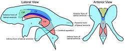 Difference between cavum septi pellucidi (CSP), cavum Vergae (CV), and cavum veli interpositi (CVI). 3=third ventricle, 4=fourth ventricle. | |
| Anatomical terms of neuroanatomy |
Axial MR/CT show a triangular-shaped cerebrospinal fluid (CSF) space between the lateral ventricles. On sagittal images, CVI can appear as a slit-like, linear-to-round/ovoid CSF collection below the fornices, and above the 3rd ventricle.
There are usually no associated abnormalities, although larger lesions may cause an obstructive hydrocephalus. No treatment is usually necessary.[1]
See also
References
- Emedicine -- Cavum vergae
This article is issued from
Wikipedia.
The text is licensed under Creative
Commons - Attribution - Sharealike.
Additional terms may apply for the media files.