Carotid-cavernous fistula
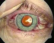
A carotid-cavernous fistula results from an abnormal communication between the arterial and venous systems within the cavernous sinus in the skull. It is a type of arteriovenous fistula. As arterial blood under high pressure enters the cavernous sinus, the normal venous return to the cavernous sinus is impeded and this causes engorgement of the draining veins, manifesting most dramatically as a sudden engorgement and redness of the eye of the same side.
| Carotid-cavernous fistula | |
|---|---|
| Other names | CCF |
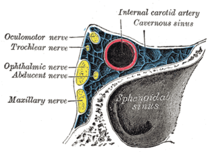 | |
| Oblique section through the cavernous sinus. | |
| Specialty | Neurology, cardiology |
Presentation
CCF symptoms include bruit (a humming sound within the skull due to high blood flow through the arteriovenous fistula), progressive visual loss, and pulsatile proptosis or progressive bulging of the eye due to dilatation of the veins draining the eye. Pain is the symptom that patients often find the most difficult to tolerate.
Patients usually present with sudden or insidious onset of redness in one eye, associated with progressive proptosis or bulging.
They may have a history of similar episodes in the past.
Causes
Carotid cavernous fistulae may form following closed or penetrating head trauma, surgical damage, rupture of an intracavernous aneurysm, or in association with connective tissue disorders, vascular diseases and dural fistulas.
Diagnosis
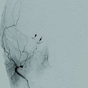
This is based on MRI scan, magnetic resonance angiography and CT scan. A cerebral digital subtraction angiography (DSA) enhances visualization of the fistula.
- CT scans classically show an enlarged superior ophthalmic vein, cavernous sinus enlargement ipsilateral (same side) as the abnormality and possibly diffuse enlargement of all the extraocular muscles resulting from venous engorgement.
- Selective arteriography is used to evaluate arteriovenous fistulas.
- High resolution digital subtraction angiography may help in classifying CCF into dural and direct type and thus formulate a strategy to treat it either by a balloon or coil or both with or without preservation of parent ipsilateral carotid artery.[2]
Classification
Various classifications have been proposed for CCF. They may be divided into low-flow or high-flow, traumatic or spontaneous and direct or indirect. The traumatic CCF typically occurs after a basal skull fracture. The spontaneous dural cavernous fistula which is more common usually results from a degenerative process in older patients with systemic hypertension and atherosclerosis. Direct fistulas occur when the Internal Carotid artery (ICA) itself fistulizes into the Cavernous sinus whereas indirect is when a branch of the ICA or External Carotid artery (ECA) communicates with the cavernous sinus.
A popular classification divides CCF into four varieties depending on the type of arterial supply.
| Type | Description |
|---|---|
| A | Fistulous supply from the internal carotid artery |
| B | Supply from the dural branches of internal carotid artery |
| C | from meningeal branches of ext carotid artery |
| D | combined ICA+ECA |
Treatment
The mainstay of treatment for CCF is endovascular therapy. This may be transarterial (mostly in the case of direct CCF) or transvenous (most commonly in indirect CCF). Occasionally, more direct approaches, such as direct transorbital puncture of the cavernous sinus or cannulation of the draining superior orbital vein are used when conventional approaches are not possible. Spontaneous resolution of indirect fistulae has been reported but is uncommon. Staged manual compression of the ipsilateral carotid has been reported to assist with spontaneous closure in selected cases.
Direct CCF may be treated by occlusion of the affected cavernous sinus (coils, balloon, liquid agents), or by reconstruction of the damaged internal carotid artery (stent, coils or liquid agents).
Indirect CCF may be treated by occlusion of the affected cavernous sinus with coils, liquid agents or a combination of both.[3][4][5]
References
- Thinda, S; Melson, MR; Kuchtey, RW (Jul 28, 2012). "Worsening angle closure glaucoma and choroidal detachments subsequent to closure of a carotid cavernous fistula". BMC Ophthalmology. 12: 28. doi:10.1186/1471-2415-12-28. PMC 3412712. PMID 22839357.
- Dr Bharatkumar Mudalgi, Dr Manohar Kachare, Dr Manish Shrivastava, Dr Akshay Kulkarni, Endovascular Treatment of Carotid Cavernous Fistulae: A Study of Management and Outcome, Journal of medical science and clinical research Dr Bharatkumar Mudalgi et al JMSCR Volume 05 Issue 07 July 2017 https://dx.doi.org/10.18535/jmscr/v5i7.135
- Ong, CK; Wang, LL; Parkinson, RJ; Wenderoth, JD (2009). "Onyx embolisation of cavernous sinus dural arteriovenous fistula via direct percutaneous transorbital puncture". Journal of Medical Imaging and Radiation Oncology. 53 (3): 291–5. doi:10.1111/j.1754-9485.2009.02086.x. PMID 19624295.
- Bhatia, Kartik D; Wang, Lily; Parkinson, Richard J; Wenderoth, Jason D (2009). "Successful Treatment of Six Cases of Indirect Carotid-Cavernous Fistula with Ethylene Vinyl Alcohol Copolymer (Onyx) Transvenous Embolization". Journal of Neuro-Ophthalmology. 29 (1): 3–8. doi:10.1097/WNO.0b013e318199c85c. PMID 19458567.
- Nadarajah, M.; Power, M.; Barry, B.; Wenderoth, J. (2011). "Treatment of a traumatic carotid-cavernous fistula by the sole use of a flow diverting stent". Journal of NeuroInterventional Surgery. 4 (3): e1. doi:10.1136/neurintsurg-2011-010000. PMID 21990483.