Capsule endoscopy
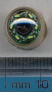
Capsule endoscopy is a procedure used to record internal images of the gastrointestinal tract for use in medical diagnosis. Newer developments are also able to take biopsies and release medication at specific locations of the entire gastrointestinal tract . The capsule (aka pill cam) is similar in shape to a standard pharmaceutical capsule, although a little larger, and contains a tiny camera and an array of LEDs powered by a battery. After a patient swallows the capsule, it passes along the gastrointestinal tract taking a number of images per second which are transmitted wirelessly to an array of receivers connected to a portable recording device carried by the patient. The primary use of capsule endoscopy is to examine areas of the small intestine that cannot be seen by other types of endoscopy such as colonoscopy or esophagogastroduodenoscopy (EGD).[1]
| Wireless Capsule Endoscopy | |
|---|---|
| Medical diagnostics | |
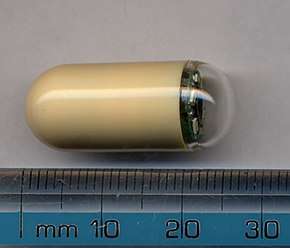 Picture of a capsule | |
| MeSH | D053704 |
| OPS-301 code | 1-63a, 1-656 |
Medical uses
Esophagogastroduodenoscopy (EGD), employs a camera attached to a long flexible tube to view the upper portion of the gastrointestinal tract, namely the esophagus, the stomach and the beginning of the first part of the small intestine called the duodenum, and a colonoscope, inserted through the rectum, can view the colon and the distal portion of the small intestine, the terminal ileum, however, these two types of endoscopy cannot visualize the majority of the middle portion of the small intestine. Capsule endoscopy is used to examine parts of the gastrointestinal tract that cannot be seen with other types of endoscopy. It is useful when disease is suspected in the small intestine, and can sometimes be used to find the site of gastrointestinal bleeding or the cause of unexplained abdominal pain, such as Crohn's disease. However, unlike EGD or colonoscopy it cannot be used to treat pathology that may be discovered. Common reasons for using capsule endoscopy include diagnosis of unexplained bleeding, iron deficiency, or abdominal pain, searching for polyps, ulcers and tumors of small intestine, and diagnosis of inflammatory bowel disease.[2] The images collected by the miniature camera during a session are transferred wirelessly to an external receiver worn by the patient, using any one of a band of appropriate frequencies. The collected images are then transferred to a computer for display, review and diagnosis.[3] A transmitted radio-frequency signal can be used to accurately estimate the location of the capsule and to track it in real time inside the body and gastrointestinal tract.[4] It is unclear if capsule endoscopy can replace gastroscopy for those with cirrhosis.[5]
As of 2014 research was targeting additional sensing mechanisms and localization and motion control systems to enable new applications for the technology, for example, drug delivery. Wireless energy transmission was also being investigated as a way of providing a continuous energy source for the capsule.[6]
Manufacturers
As of 2015 there were a number of manufacturers.[7] The technology was originally developed by Gabi Iddan and Paul Swain, with the first pill swallowed in 1997;[8] Iddan founded Given Imaging which received FDA approval in 2001.[8]
Side effects
Capsule endoscopy is considered to be a very safe method to determine an unknown cause of a gastrointestinal bleed.[9] The capsule is usually excreted with the feces within 24–48 hours.[9] There has been a report of retention of the capsule for almost four and a half years although the patient was asymptomatic.[9] However, the risk of bowel obstruction may be countered by abdominal X-ray to locate the device for removal by endoscopy or surgery.[9]
Risk of retention
In a review of 22,840 cases, the capsule was retained 1.4% of the time, with Crohn's disease a common cause; most were surgically removed.[10]
External links
- Macrae, Fiona (7 August 2014). "Camera takes 18 photos per second while travelling through your body". Mail Online. The article includes a video of the gastrointestinal tract imaged by capsule endoscopy.
References
- "The endoscopy procedures we provide". 28 November 2018. Farzan Bahin, The Hills Gastroenterology, Sydney, Australia.
- Tong J, Svarta S, Ou G, Kwok R, Law J, Enns R (October 2012). "Diagnostic yield of capsule endoscopy in the setting of iron deficiency anemia without evidence of gastrointestinal bleeding". Canadian Journal of Gastroenterology. 26 (10): 687–90. doi:10.1155/2012/182542. PMC 3472906. PMID 23061059.
- Yuce MR, Dissanayake T (2012). "Easy-to-swallow wireless telemetry". IEEE Microwave Magazine. 13 (6): 90–101. doi:10.1109/MMM.2012.2205833.
- Pourhomayoun M, Fowler ML, Jin Z (2012). "A Novel Method for Medical Implant In-Body Localization" (PDF). Proceedings of the IEEE International Conference of Engineering in Medicine and Biology.
- Colli A, Gana JC, Turner D, Yap J, Adams-Webber T, Ling SC, Casazza G (October 2014). "Capsule endoscopy for the diagnosis of oesophageal varices in people with chronic liver disease or portal vein thrombosis" (PDF). The Cochrane Database of Systematic Reviews. 10 (10): CD008760. doi:10.1002/14651858.CD008760.pub2. PMID 25271409.
- Yuce MR, Alici G, Than TD (2014). Wireless Endoscopy. Wiley Encyclopedia of Electrical and Electronics Engineering. pp. 1–25. doi:10.1002/047134608X.W8233. ISBN 9780471346081.
- Slawinski PR, Obstein KL, Valdastri P (October 2015). "Capsule endoscopy of the future: What's on the horizon?". World Journal of Gastroenterology. 21 (37): 10528–41. doi:10.3748/wjg.v21.i37.10528. PMC 4588075. PMID 26457013.
- Adler SN, Metzger YC (July 2011). "PillCam COLON capsule endoscopy: recent advances and new insights". Therapeutic Advances in Gastroenterology. 4 (4): 265–8. doi:10.1177/1756283X11401645. PMC 3131168. PMID 21765870.
- Bhattarai M, Bansal P, Khan Y (July 2013). "Longest duration of retention of video capsule: A case report and literature review". World Journal of Gastrointestinal Endoscopy. 5 (7): 352–5. doi:10.4253/wjge.v5.i7.352. PMC 3711067. PMID 23858380.
- Liao Z, Gao R, Xu C, Li ZS (February 2010). "Indications and detection, completion, and retention rates of small-bowel capsule endoscopy: a systematic review". Gastrointestinal Endoscopy. 71 (2): 280–6. doi:10.1016/j.gie.2009.09.031. PMID 20152309.