Calcifying odontogenic cyst
Calcifying odotogenic cyst is a benign odontogenic tumor of cystic type most likely to affect the anterior areas of the jaws. It is most common in people in their second to third decades but can be seen at almost any age. On radiographs, the calcifying odontogenic cyst appears as a unilocular radiolucency (dark area). In one-third of cases, an impacted tooth is involved. Microscopically, there are many cells that are described as "ghost cells", enlarged eosinophilic epithelial cells without nuclei.
| Calcifying odontogenic cyst | |
|---|---|
| Other names | Gorlin cyst, calcifying cystic odontogenic tumor[1] |
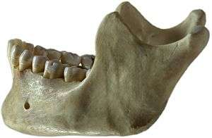 | |
| This condition usually affects the jaw area | |
| Specialty | Dentistry |
Signs and symptoms
It is a rare type of cyst and can occur at any age, mostly between second and third decade of life. The diameter ranges from 2 to 4 cm and swelling pain may be present. Intrabony expansions may produce hard bony expansion and may perforate cortical bones. It can also extend to soft tissue. It can present asymptomaticaly.[2]
Pathogenesis
Epithelial lining has ability to induce formation of dental tissues in adjacent connective tissue wall.[2]
Diagnosis
Radiographic features
Unilocular, multilocular or mixed radiolucency can be seen. Irregular calcifications may be seen in some cases.
Histology
In general, the epithelium seen is of stratified squamous type and is 5–8 cells thick. Additionally, focal areas of stellate reticulum like cells are seen and near the basement membrane ameloblast-like cells may be seen. Each type of calcifying odontogenic cyst shows special features of which there are three types:
1)Type 1A. Ghost cells and dentinoid are seen.
2)Type 1B. Formation of calcified tissues in the lumen of the cyst wall showing dystrophic calcification. Proliferation of tissues is similar to an Ameloblastic Fibroma.
3)Type 1C. Ameloblast-like proliferation in the connective tissue and lumen of the cyst may be seen.
Treatment
The standard treatment of calcifying odontogenic cyst is enucleation and curettage. Recurrence following enucleation and curretage is rare.[3]
See also
References
- Kler, Shikha; Palaskar, Sangeeta; Shetty, and, Vishwa Parkash; Bhushan, Anju (2009), "Intraosseous calcifying cystic odontogenic tumor", J Oral Maxillofac Pathol, 13 (1): 27–29, doi:10.4103/0973-029X.48753, PMC 3162852, PMID 21886994.
- Regezi JA, Sciubba J, Jordan RCK (2012-04-15). Oral Pathology: Clinical Pathologic Correlations. Elsevier Health Sciences. p. 259. ISBN 1-4557-0269-2.
- Mervyn Shear; Paul Speight (2008-04-15). Cysts of the Oral and Maxillofacial Regions. John Wiley & Sons. p. 107. ISBN 978-0-470-75972-1.