Bulbospongiosus muscle
The bulbospongiosus muscle (bulbocavernosus in older texts) is one of the superficial muscles of the perineum.[1] It has a slightly different origin, insertion and function in males and females. In males, it covers the bulb of the penis. In females, it covers the vestibular bulb.
| Bulbospongiosus muscle | |
|---|---|
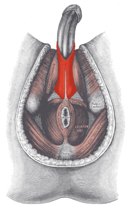 Muscles of male perineum. (Bulbocavernosus visible at upper left.) | |
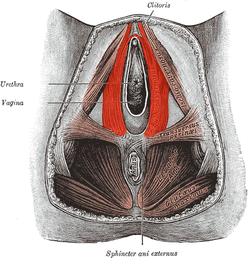 Muscles of the female perineum. (Bulbocavernosus visible at upper right.) | |
| Details | |
| Origin | Median raphe |
| Artery | Perineal artery |
| Nerve | pudendal nerve |
| Actions | in males, empties the urethra; in females, clenches the vagina |
| Identifiers | |
| Latin | musculus bulbospongiosus |
| TA | A09.5.02.005 |
| FMA | 19729 |
| Anatomical terms of muscle | |
In both sexes, it is innervated by the deep/muscular branch of the perineal nerve, which is a branch of the pudendal nerve.
Structure
In males, the bulbospongiosus is located in the middle line of the perineum, in front of the anus. It consists of two symmetrical parts, united along the median line by a tendinous perineal raphe. It arises from the central tendinous point of the perineum and from the median perineal raphe in front.
In females, there is no union, nor a tendinous perineal raphe; the parts are disjoint primarily and arise from the same central tendinous point of the perinium, which is the tendon that is formed at the point where the bulbospongiosus muscle, superficial transverse perineal muscle, and external anal sphincter muscle converge to form this major supportive structure of vagina and other organs, and from the clitoris in front.[2]
Fibers
Its fibers diverge; the most posterior form a thin layer, which is lost on the inferior fascia of the urogenital diaphragm; the middle fibers encircle the bulb and adjacent parts, of the corpus cavernosum urethrae, and join with the fibers of the opposite side, on the upper part of the corpus cavernosum urethrae, in a strong aponeurosis; the anterior fibers, spread out over the side of the corpus cavernosum penis, to be inserted partly into that body, anterior to the Ischiocavernosus, occasionally extending to the pubis, and partly ending in a tendinous expansion which covers the dorsal vessels of the penis.
The latter fibers are best seen by dividing the muscle longitudinally, and reflecting it from the surface of the corpus cavernosum urethra.
Function
In males it contributes to erection, the contractions of orgasm and ejaculation.[3] In females it contributes to clitoral erection and the contractions of orgasm, and closes the vagina.
This muscle serves to empty the canal of the urethra, after the bladder has expelled its contents; during the greater part of the act of micturition its fibers are relaxed, and it only comes into action at the end of the process.
The middle fibers are supposed by Krause to assist in the erection of the corpus spongiosum, by compressing the erectile tissue of the bulb.
The anterior fibers also contribute to the erection of the penis by compressing the deep dorsal vein of the penis as they are inserted into, and continuous with, the fascia of the penis.
Gallery
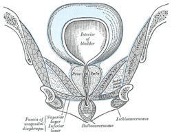 Coronal section of anterior part of the male pelvis, through the pubic arch. Seen from in front.
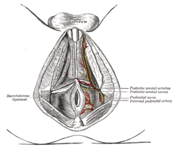 The superficial branches of the internal pudendal artery in the male.
References
- Maclean, Allan; Reid, Wendy (2011). "40". In Shaw, Robert (ed.). Gynaecology. Edinburgh New York: Churchill Livingstone/Elsevier. pp. 599–612. ISBN 978-0-7020-3120-5; Access provided by the University of Pittsburgh
- "Human Anatomy, The Female Perineum, Muscles of the Superficial Perineal Pouch". act.downstate.edu. SUNY Downstate Medical Center. Retrieved 2018-02-03.
- Marieb, Elaine (2013). Anatomy & physiology : books a la carte edition. Benjamin-Cummings. p. 895. ISBN 9780321887603.
- This article incorporates text in the public domain from page 428 of the 20th edition of Gray's Anatomy (1918)
External links
| Wikimedia Commons has media related to Bulbospongiosus muscle. |
- Anatomy figure: 42:04-02 at Human Anatomy Online, SUNY Downstate Medical Center—"Muscles of the male superficial perineal pouch."
- Anatomy photo:41:11-0102 at the SUNY Downstate Medical Center—"The Female Perineum: Muscles of the Superficial Perineal Pouch"
- Anatomy figure: 43:04-11 at Human Anatomy Online, SUNY Downstate Medical Center—"The urinary bladder and the urethra as seen in a frontal section of the female pelvis."
- Anatomy image:9162 at the SUNY Downstate Medical Center
- Anatomy image:9197 at the SUNY Downstate Medical Center
| Authority control |
|---|