Bone fracture
A bone fracture (sometimes abbreviated FRX or Fx, Fx, or #) is a medical condition in which there is a partial or complete break in the continuity of the bone. In more severe cases, the bone may be broken into several pieces.[1] A bone fracture may be the result of high force impact or stress, or a minimal trauma injury as a result of certain medical conditions that weaken the bones, such as osteoporosis, osteopenia, bone cancer, or osteogenesis imperfecta, where the fracture is then properly termed a pathologic fracture.[2]
| Bone fracture | |
|---|---|
| Other names | broken bone, bone break |
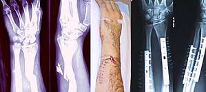 | |
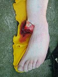
| Internal and external views of an arm with a compound fracture, both before and after surgery | |
| Specialty | Orthopedics |
Signs and symptoms
Although bone tissue itself contains no nociceptors, bone fracture is painful for several reasons:[3]
- Breaking in the continuity of the periosteum, with or without similar discontinuity in endosteum, as both contain multiple pain receptors.
- Edema and hematoma of nearby soft tissues caused by ruptured bone marrow evokes pressure pain.
- Involuntary muscle spasms trying to hold bone fragments in place.
Damage to adjacent structures such as nerves, muscles or blood vessels, spinal cord, and nerve roots (for spine fractures), or cranial contents (for skull fractures) may cause other specific signs and symptoms.
Complications
Some fractures may lead to serious complications including a condition known as compartment syndrome. If not treated, eventually, compartment syndrome may require amputation of the affected limb. Other complications may include non-union, where the fractured bone fails to heal or mal-union, where the fractured bone heals in a deformed manner. One form of malunion is the malrotation of a bone, which is especially common after femoral and tibial fractures.
Complications of fractures may be classified into three broad groups, depending upon their time of occurrence. These are as follows –
- Immediate complications – occurs at the time of the fracture.
- Early complications – occurring in the initial few days after the fracture.
- Late complications – occurring a long time after the fracture.
| Immediate complications | Early complications | Late complications |
|---|---|---|
Systemic
|
Systemic
|
Imperfect union of the fracture
|
Local
|
Local
|
Others
|
Pathophysiology
The natural process of healing a fracture starts when the injured bone and surrounding tissues bleed, forming a fracture hematoma. The blood coagulates to form a blood clot situated between the broken fragments. Within a few days, blood vessels grow into the jelly-like matrix of the blood clot. The new blood vessels bring phagocytes to the area, which gradually removes the non-viable material. The blood vessels also bring fibroblasts in the walls of the vessels and these multiply and produce collagen fibres. In this way, the blood clot is replaced by a matrix of collagen. Collagen's rubbery consistency allows bone fragments to move only a small amount unless severe or persistent force is applied.
At this stage, some of the fibroblasts begin to lay down bone matrix in the form of collagen monomers. These monomers spontaneously assemble to form the bone matrix, for which bone crystals (calcium hydroxyapatite) are deposited in amongst, in the form of insoluble crystals. This mineralization of the collagen matrix stiffens it and transforms it into bone. In fact, bone is a mineralized collagen matrix; if the mineral is dissolved out of bone, it becomes rubbery. Healing bone callus on average is sufficiently mineralized to show up on X-ray within 6 weeks in adults and less in children. This initial "woven" bone does not have the strong mechanical properties of mature bone. By a process of remodelling, the woven bone is replaced by mature "lamellar" bone. The whole process may take up to 18 months, but in adults, the strength of the healing bone is usually 80% of normal by 3 months after the injury.
Several factors may help or hinder the bone healing process. For example, tobacco smoking hinders the process of bone healing,[4] and adequate nutrition (including calcium intake) will help the bone healing process. Weight-bearing stress on bone, after the bone has healed sufficiently to bear the weight, also builds bone strength.
Although there are theoretical concerns about NSAIDs slowing the rate of healing, there is not enough evidence to warrant withholding the use of this type analgesic in simple fractures.[5]
Effects of smoking
Smokers generally have lower bone density than non-smokers, so they have a much higher risk of fractures. There is also evidence that smoking delays bone healing.[6]
Diagnosis
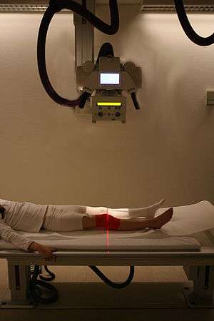
A bone fracture may be diagnosed based on the history given and the physical examination performed. Radiographic imaging often is performed to confirm the diagnosis. Under certain circumstances, radiographic examination of the nearby joints is indicated in order to exclude dislocations and fracture-dislocations. In situations where projectional radiography alone is insufficient, Computed Tomography (CT) or Magnetic Resonance Imaging (MRI) may be indicated.
Classification
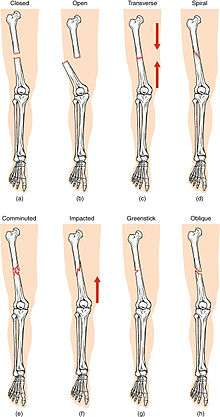
(a) closed fracture
(b) open fracture
(c) transverse fracture
(d) spiral fracture
(e) comminuted fracture
(f) impacted fracture
(g) greenstick fracture
(h) oblique fracture
In orthopedic medicine, fractures are classified in various ways. Historically they are named after the physician who first described the fracture conditions, however, there are more systematic classifications as well.
They may be divided into stable versus unstable depending on the likelihood that they may shift further.
Mechanism
- Traumatic fracture – This is a fracture due to sustained trauma. e.g., fractures caused by a fall, road traffic accident, fight, etc.
- Pathologic fracture – A fracture through a bone that has been made weak by some underlying disease is called pathological fracture. e.g., a fracture through a bone weakened by metastasis. Osteoporosis is the most common cause of pathological fracture.
- Periprosthetic fracture – This is a fracture at the point of mechanical weakness at the end of an implant
Soft-tissue involvement
- Closed fractures are those in which the overlying skin is intact
- Open/compound fractures involve wounds that communicate with the fracture, or where fracture hematoma is exposed, and may thus expose bone to contamination. Open injuries carry a higher risk of infection.
- Clean fracture
- Contaminated fracture
Displacement
Fracture pattern
- Linear fracture: A fracture that is parallel to the bone's long axis
- Transverse fracture: A fracture that is at a right angle to the bone's long axis
- Oblique fracture: A fracture that is diagonal to a bone's long axis (more than 30°)
- Spiral fracture: A fracture where at least one part of the bone has been twisted
- Compression fracture/wedge fracture: usually occurs in the vertebrae, for example when the front portion of a vertebra in the spine collapses due to osteoporosis (a medical condition which causes bones to become brittle and susceptible to fracture, with or without trauma)
- Impacted fracture: A fracture caused when bone fragments are driven into each other
- Avulsion fracture: A fracture where a fragment of bone is separated from the main mass
Fragments
- Incomplete fracture: Is a fracture in which the bone fragments are still partially joined, in such cases, there is a crack in the osseous tissue that does not completely traverse the width of the bone.
- Complete fracture: Is a fracture in which bone fragments separate completely.
- Comminuted fracture: Is a fracture in which the bone has broken into several pieces.
Anatomical location
An anatomical classification may begin with specifying the involved body part, such as the head or arm, followed with more specific localization. Fractures that have additional definition criteria than merely localization often may be classified as subtypes of fractures, such as a Holstein-Lewis fracture being a subtype of a humerus fracture. Most typical examples in an orthopaedic classification given in the previous section cannot be classified appropriately into any specific part of an anatomical classification, however, as they may apply to multiple anatomical fracture sites.
- Skull fracture
- Basilar skull fracture
- Blowout fracture – a fracture of the walls or floor of the orbit
- Mandibular fracture
- Nasal fracture
- Le Fort fracture of skull – facial fractures involving the maxillary bone and surrounding structures in a usually bilateral and either horizontal, pyramidal, or transverse way.
- Spinal fracture
- Cervical fracture
- Fracture of C1, including Jefferson fracture
- Fracture of C2, including Hangman's fracture
- Flexion teardrop fracture – a fracture of the anteroinferior aspect of a cervical vertebral
- Clay-shoveler fracture – fracture through the spinous process of a vertebra occurring at any of the lower cervical or upper thoracic vertebrae
- Burst fracture – in which a vertebra breaks from a high-energy axial load
- Compression fracture – a collapse of a vertebra, often in the form of wedge fractures due to larger compression anteriorly
- Chance fracture – compression injury to the anterior portion of a vertebral body with concomitant distraction injury to posterior elements
- Holdsworth fracture – an unstable fracture dislocation of the thoracolumbar junction of the spine
- Cervical fracture
- Rib fracture
- Sternal fracture
- Shoulder fracture
- Clavicle fracture
- Scapular fracture
- Arm fracture
- Humerus fracture (fracture of upper arm)
- Supracondylar fracture
- Holstein-Lewis fracture – a fracture of the distal third of the humerus resulting in entrapment of the radial nerve
- Forearm fracture
- Ulnar fracture
- Monteggia fracture – a fracture of the proximal third of the ulna with the dislocation of the head of the radius
- Hume fracture – a fracture of the olecranon with an associated anterior dislocation of the radial head
- Radius fracture
- Essex-Lopresti fracture – a fracture of the radial head with concomitant dislocation of the distal radio-ulnar joint with disruption of the interosseous membrane [8]
- Distal radius fracture
- Galeazzi fracture – a fracture of the radius with dislocation of the distal radioulnar joint
- Colles' fracture – a distal fracture of the radius with dorsal (posterior) displacement of the wrist and hand
- Smith's fracture – a distal fracture of the radius with volar (ventral) displacement of the wrist and hand
- Barton's fracture – an intra-articular fracture of the distal radius with dislocation of the radiocarpal joint
- Ulnar fracture
- Humerus fracture (fracture of upper arm)
- Hand fracture
- Scaphoid fracture
- Rolando fracture – a comminuted intra-articular fracture through the base of the first metacarpal bone
- Bennett's fracture – a fracture of the base of the first metacarpal bone which extends into the carpometacarpal (CMC) joint [9]
- Boxer's fracture – a fracture at the neck of a metacarpal
- Pelvic fracture
- Fracture of the hip bone
- Duverney fracture – an isolated pelvic fracture involving only the iliac wing
- Femoral fracture
- Hip fracture (anatomically a fracture of the femur bone and not the hip bone)
- Patella fracture
- Crus fracture
- Tibia fracture
- Pilon fracture
- Tibial plateau fracture
- Bumper fracture – a fracture of the lateral tibial plateau caused by a forced valgus applied to the knee
- Segond fracture – an avulsion fracture of the lateral tibial condyle
- Gosselin fracture – a fractures of the tibial plafond into anterior and posterior fragments [10]
- Toddler's fracture – an undisplaced and spiral fracture of the distal third to distal half of the tibia [11]
- Fibular fracture
- Maisonneuve fracture – a spiral fracture of the proximal third of the fibula associated with a tear of the distal tibiofibular syndesmosis and the interosseous membrane
- Le Fort fracture of ankle – a vertical fracture of the antero-medial part of the distal fibula with avulsion of the anterior tibiofibular ligament [10]
- Bosworth fracture – a fracture with an associated fixed posterior dislocation of the distal fibular fragment that becomes trapped behind the posterior tibial tubercle; the injury is caused by severe external rotation of the ankle [12]
- Combined tibia and fibula fracture
- Trimalleolar fracture – involving the lateral malleolus, medial malleolus, and the distal posterior aspect of the tibia
- Bimalleolar fracture – involving the lateral malleolus and the medial malleolus
- Pott's fracture
- Tibia fracture
- Foot fracture
- Lisfranc fracture – in which one or all of the metatarsals are displaced from the tarsus[13]
- Jones fracture – a fracture of the proximal end of the fifth metatarsal
- March fracture – a fracture of the distal third of one of the metatarsals occurring because of recurrent stress
- Calcaneal fracture - a fracture of the calcaneus (heel bone)
OTA/AO classification
The Orthopaedic Trauma Association Committee for Coding and Classification published its classification system [14] in 1996, adopting a similar system to the 1987 AO Foundation system.[15] In 2007, they extended their system,[16] unifying the two systems regarding wrist, hand, foot, and ankle fractures.
Classifications named after people
A number of classifications are named after the person (eponymous) who developed it.
- "Denis classification" for spinal fractures [17]
- "Frykman classification" for forearm fractures (fractures of radius and ulna)
- "Gustilo open fracture classification" [18]
- "Letournel and Judet Classification" for Acetabular fractures [19]
- "Neer classification" for humerus fractures [20][21]
- Seinsheimer classification, Evans-Jensen classification, Pipkin classification, and Garden classification for hip fractures
Prevention
Both high- and low-force trauma can cause bone fracture injuries.[22][23] Preventive efforts to reduce motor vehicle crashes, the most common cause of high-force trauma, include reducing distractions while driving.[24] Common distractions are driving under the influence and texting or calling while driving, both of which lead to an approximate 6-fold increase in crashes.[24] Wearing a seatbelt can also reduce the likelihood of injury in a collision.[24]
A common cause of low-force trauma is an at-home fall.[22][23] When considering preventative efforts, the National Institute of Health (NIH) examines ways to reduce the likelihood of falling, the force of the fall, and bone fragility.[25] To prevent at-home falls they suggest keeping cords out of high-traffic areas where someone could trip, installing handrails and keeping stairways well-lit, and installing an assistive bar near the bathtub in the washroom for support.[25] To reduce the impact of a fall the NIH recommends to try falling straight down on your buttocks or onto your hands.[25] Finally, taking calcium vitamin D supplements can help strengthen your bones.[25]
Treatment
Treatment of bone fractures are broadly classified as surgical or conservative, the latter basically referring to any non-surgical procedure, such as pain management, immobilization or other non-surgical stabilization. A similar classification is open versus closed treatment, in which open treatment refers to any treatment in which the fracture site is opened surgically, regardless of whether the fracture is an open or closed fracture.
Pain management
In arm fractures in children, ibuprofen has been found to be as effective as a combination of acetaminophen and codeine.[26]
Immobilization
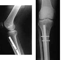
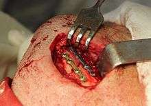
Since bone healing is a natural process that will occur most often, fracture treatment aims to ensure the best possible function of the injured part after healing. Bone fractures typically are treated by restoring the fractured pieces of bone to their natural positions (if necessary), and maintaining those positions while the bone heals. Often, aligning the bone, called reduction, in a good position and verifying the improved alignment with an X-ray is all that is needed. This process is extremely painful without anaesthesia, about as painful as breaking the bone itself. To this end, a fractured limb usually is immobilized with a plaster or fibreglass cast or splint that holds the bones in position and immobilizes the joints above and below the fracture. When the initial post-fracture oedema or swelling goes down, the fracture may be placed in a removable brace or orthosis. If being treated with surgery, surgical nails, screws, plates, and wires are used to hold the fractured bone together more directly. Alternatively, fractured bones may be treated by the Ilizarov method which is a form of an external fixator.
Occasionally smaller bones, such as phalanges of the toes and fingers, may be treated without the cast, by buddy wrapping them, which serves a similar function to making a cast. A device called a Suzuki frame may be used in cases of deep, complex intra-articular digit fractures.[27] By allowing only limited movement, immobilization helps preserve anatomical alignment while enabling callus formation, toward the target of achieving union.
Splinting results in the same outcome as casting in children who have a distal radius fracture with little shifting.[28]
Surgery
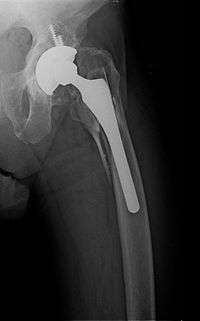
Surgical methods of treating fractures have their own risks and benefits, but usually surgery is performed only if conservative treatment has failed, is very likely to fail, or likely to result in a poor functional outcome. With some fractures such as hip fractures (usually caused by osteoporosis), surgery is offered routinely because non-operative treatment results in prolonged immobilisation, which commonly results in complications including chest infections, pressure sores, deconditioning, deep vein thrombosis (DVT), and pulmonary embolism, which are more dangerous than surgery. When a joint surface is damaged by a fracture, surgery is also commonly recommended to make an accurate anatomical reduction and restore the smoothness of the joint.
Infection is especially dangerous in bones, due to the recrudescent nature of bone infections. Bone tissue is predominantly extracellular matrix, rather than living cells, and the few blood vessels needed to support this low metabolism are only able to bring a limited number of immune cells to an injury to fight infection. For this reason, open fractures and osteotomies call for very careful antiseptic procedures and prophylactic use of antibiotics.
Occasionally, bone grafting is used to treat a fracture.
Sometimes bones are reinforced with metal. These implants must be designed and installed with care. Stress shielding occurs when plates or screws carry too large of a portion of the bone's load, causing atrophy. This problem is reduced, but not eliminated, by the use of low-modulus materials, including titanium and its alloys. The heat generated by the friction of installing hardware can accumulate easily and damage bone tissue, reducing the strength of the connections. If dissimilar metals are installed in contact with one another (i.e., a titanium plate with cobalt-chromium alloy or stainless steel screws), galvanic corrosion will result. The metal ions produced can damage the bone locally and may cause systemic effects as well.
Other
A Cochrane review of low-intensity pulsed ultrasound to speed healing in newly broken bones found insufficient evidence to justify routine use.[29] Other reviews have found tentative evidence of benefit.[30] It may be an alternative to surgery for established nonunions.[31]
Vitamin D supplements combined with additional calcium marginally reduces the risk of hip fractures and other types of fracture in older adults; however, vitamin D supplementation alone did not reduce the risk of fractures.[32]
Children
In children, whose bones are still developing, there are risks of either a growth plate injury or a greenstick fracture.
- A greenstick fracture occurs due to mechanical failure on the tension side. That is since the bone is not so brittle as it would be in an adult, it does not completely fracture, but rather exhibits bowing without complete disruption of the bone's cortex in the surface opposite the applied force.
- Growth plate injuries, as in Salter-Harris fractures, require careful treatment and accurate reduction to make sure that the bone continues to grow normally.
- Plastic deformation of the bone, in which the bone permanently bends, but does not break, also is possible in children. These injuries may require an osteotomy (bone cut) to realign the bone if it is fixed and cannot be realigned by closed methods.
- Certain fractures mainly occur in children, including fracture of the clavicle and supracondylar fracture of the humerus.
See also
- Stress fracture
- Distraction osteogenesis
- Rickets
- Catagmatic
- H. Winnett Orr, U.S. Army surgeon who developed Orthopedic plaster casts
References
- Katherine, Abel (2013). Official CPC Certification Study Guide. American Medical Association. p. 108.
- Witmer, Daniel K.; Marshall, Silas T.; Browner, Bruce D. (2016). "Emergency Care of Musculoskeletal Injuries". In Townsend, Courtney M.; Beauchamp, R. Daniel; Evers, B. Mark; Mattox, Kenneth L. (eds.). Sabiston Textbook of Surgery (20th ed.). Elsevier. pp. 462–504. ISBN 978-0-323-40163-0.
- MedicineNet – Fracture Archived 2008-12-21 at the Wayback Machine Medical Author: Benjamin C. Wedro, MD, FAAEM.
- Sloan, A.; Hussain, I.; Maqsood, M.; Eremin, O.; El-Sheemy, M. (2010). "The effects of smoking on fracture healing". The Surgeon. 8 (2): 111–6. doi:10.1016/j.surge.2009.10.014. PMID 20303894.
- Pountos, Ippokratis; Georgouli, Theodora; Calori, Giorgio M.; Giannoudis, Peter V. (2012). "Do Nonsteroidal Anti-Inflammatory Drugs Affect Bone Healing? A Critical Analysis". The Scientific World Journal. 2012: 1–14. doi:10.1100/2012/606404. PMC 3259713. PMID 22272177.
- Kanis, J. A.; Johnell, O.; Oden, A.; Johansson, H.; De Laet, C.; Eisman, J. A.; Fujiwara, S.; Kroger, H.; McCloskey, E. V.; Mellstrom, D.; Melton, L. J.; Pols, H.; Reeve, J.; Silman, A.; Tenenhouse, A. (2004). "Smoking and fracture risk: A meta-analysis". Osteoporosis International. 16 (2): 155–62. doi:10.1007/s00198-004-1640-3. PMID 15175845.
- Roberto Schubert. "Fractures of the extremities (general rules and nomenclature)". Radiopaedia. Retrieved 2018-02-21.
- Essex Lopresti fracture Archived 2009-10-01 at the Wayback Machine at Wheeless' Textbook of Orthopaedics online
- "Bennett's fracture-subluxation". GPnotebook.
- Hunter, Tim B.; Peltier, Leonard F.; Lund, Pamela J. (2000). "Radiologic History Exhibit". RadioGraphics. 20 (3): 819–36. doi:10.1148/radiographics.20.3.g00ma20819. PMID 10835130.
- Mellick, Larry B.; Milker, Laura; Egsieker, Erik (1999). "Childhood accidental spiral tibial (CAST) fractures". Pediatric Emergency Care. 15 (5): 307–9. doi:10.1097/00006565-199910000-00001. PMID 10532655.
- Perry, C. R.; Rice, S; Rao, A; Burdge, R (1983). "Posterior fracture-dislocation of the distal part of the fibula. Mechanism and staging of injury". The Journal of Bone and Joint Surgery. American Volume. 65 (8): 1149–57. doi:10.2106/00004623-198365080-00016. PMID 6630259.
- TheFreeDictionary Lisfranc's fracture Citing: Mosby's Medical Dictionary, 8th edition. Copyright 2009
- "Fracture and dislocation compendium. Orthopaedic Trauma Association Committee for Coding and Classification". Journal of Orthopaedic Trauma. 10 Suppl 1: v–ix, 1–154. 1996. PMID 8814583.
- Müller ME, Nazarian S, Koch P (1987). Classification AO des fractures. Tome I. Les os longs. Berlin: Springer-Verlag.
- Marsh, J. L.; Slongo, T. F.; Agel, J; Broderick, J. S.; Creevey, W; Decoster, T. A.; Prokuski, L; Sirkin, M. S.; Ziran, B; Henley, B; Audigé, L (2007). "Fracture and dislocation classification compendium - 2007: Orthopaedic Trauma Association classification, database and outcomes committee". Journal of Orthopaedic Trauma. 21 (10 Suppl): S1–133. doi:10.1097/00005131-200711101-00001. PMID 18277234.
- "Denis classification of spinal fractures". GPnotebook.
- Rüedi, etc. all; Thomas P. Rüedi; Richard E. Buckley; Christopher G. Moran (2007). AO principles of fracture management, Volume 1. Thieme. p. 96. ISBN 978-3-13-117442-0.
- "Fractures of the Acetabulum". wheelessonline.com. Archived from the original on 2009-09-26.
- Mourad, L (1997). "Neer classification of fractures of the proximal humerus". Orthopedic Nursing. 16 (2): 76. PMID 9155417.
- Proximal Humerus Fractures at eMedicine
- "Open Fractures - OrthoInfo - AAOS". Retrieved 2018-12-03.
- Court-Brown, Charles M.; Bugler, Kate E.; Clement, Nicholas D.; Duckworth, Andrew D.; McQueen, Margaret M. (June 2012). "The epidemiology of open fractures in adults. A 15-year review". Injury. 43 (6): 891–897. doi:10.1016/j.injury.2011.12.007. ISSN 1879-0267. PMID 22204774.
- Sidwell, Richard; Matar, Maher M.; Sakran, Joseph V. (2017-10-01). "Trauma Education and Prevention". Surgical Clinics of North America. 97 (5): 1185–1197. doi:10.1016/j.suc.2017.06.010. ISSN 0039-6109. PMID 28958365.
- "Preventing Falls and Related Fractures | NIH Osteoporosis and Related Bone Diseases National Resource Center". www.bones.nih.gov. Retrieved 2018-12-03.
- Drendel, Amy L.; Gorelick, Marc H.; Weisman, Steven J.; Lyon, Roger; Brousseau, David C.; Kim, Michael K. (2009). "A Randomized Clinical Trial of Ibuprofen Versus Acetaminophen with Codeine for Acute Pediatric Arm Fracture Pain". Annals of Emergency Medicine. 54 (4): 553–60. doi:10.1016/j.annemergmed.2009.06.005. PMID 19692147.
- Keramidas EG, Miller G (October 2005). "The Suzuki frame for complex intraarticular fractures of the thumb". Plastic and Reconstructive Surgery. 116 (5): 1326–31. doi:10.1097/01.prs.0000181786.39062.0b. PMID 16217475.
- Boutis, K.; Willan, A.; Babyn, P.; Goeree, R.; Howard, A. (2010). "Cast versus splint in children with minimally angulated fractures of the distal radius: A randomized controlled trial". Canadian Medical Association Journal. 182 (14): 1507–12. doi:10.1503/cmaj.100119. PMC 2950182. PMID 20823169.
- Griffin, XL; Parsons, N; Costa, ML; Metcalfe, D (23 June 2014). "Ultrasound and shockwave therapy for acute fractures in adults". The Cochrane Database of Systematic Reviews (6): CD008579. doi:10.1002/14651858.CD008579.pub3. PMID 24956457.
- Lou, S.; Lv, H.; Li, Z.; Zhang, L.; Tang, P (1 September 2017). "The effects of low-intensity pulsed ultrasound on fresh fracture: A meta-analysis". Medince. 96 (39): e8181. doi:10.1097/MD.0000000000008181. PMC 5626319. PMID 28953676.
- Leighton, R.; Watson, J.T; Giannoudis, P.; Papakostidis, C.; Harrison, A.; Steen, R.G. (May 2017). "Healing of fracture nonunions treated with low-intensity pulsed ultrasound (LIPUS): A systematic review and meta-analysis". Injury. 48 (7): 1339–1347. doi:10.1016/j.injury.2017.05.016. PMID 28532896.

- Avenell, Alison; Mak, Jenson C. S.; O'Connell, Dianne (2014-04-14). "Vitamin D and vitamin D analogues for preventing fractures in post-menopausal women and older men". The Cochrane Database of Systematic Reviews (4): CD000227. doi:10.1002/14651858.CD000227.pub4. ISSN 1469-493X. PMID 24729336.
External links
| Classification | |
|---|---|
| External resources |
| Wikimedia Commons has media related to Bone fractures. |
- Authoritative information in orthopaedic surgery American Association of Orthopedic Surgeons (AAOS)
- Radiographic Atlas of Fracture
| Authority control |
|
|---|