Gastrulation
Gastrulation is a phase early in the embryonic development of most animals, during which the single-layered blastula is reorganized into a multilayered structure known as the gastrula. Before gastrulation, the embryo is a continuous epithelial sheet of cells; by the end of gastrulation, the embryo has begun differentiation to establish distinct cell lineages, set up the basic axes of the body (e.g. dorsal-ventral, anterior-posterior), and internalized one or more cell types including the prospective gut.
| Gastrulation | |
|---|---|
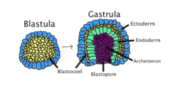 Gastrulation occurs when a blastula, made up of one layer, folds inward and enlarges to create a gastrula. This diagram is color-coded: ectoderm, blue; endoderm, green; blastocoel (the yolk sack), yellow; and archenteron (the gut), purple. | |
| Identifiers | |
| MeSH | D054262 |
| Anatomical terminology | |
In triploblastic organisms the gastrula is trilaminar ("three-layered"). These three germ layers are known as the ectoderm, mesoderm, and endoderm.[1][2] In diploblastic organisms, such as Cnidaria and Ctenophora, the gastrula has only ectoderm and endoderm. The two layers are also sometimes referred to as the hypoblast and epiblast.[3]
Gastrulation takes place after cleavage and the formation of the blastula. Gastrulation is followed by organogenesis, when individual organs develop within the newly formed germ layers.[4] Each layer gives rise to specific tissues and organs in the developing embryo. The ectoderm gives rise to epidermis, the nervous system, and to the neural crest in vertebrates. The endoderm gives rise to the epithelium of the digestive system and respiratory system, and organs associated with the digestive system, such as the liver and pancreas. The mesoderm gives rise to many cell types such as muscle, bone, and connective tissue. In vertebrates, mesoderm derivatives include the notochord, the heart, blood and blood vessels, the cartilage of the ribs and vertebrae, and the dermis.[5] Following gastrulation, cells in the body are either organized into sheets of connected cells (as in epithelia), or as a mesh of isolated cells, such as mesenchyme.[2][6]
The molecular mechanism and timing of gastrulation is different in different organisms. However, some common features of gastrulation across triploblastic organisms include: (1) A change in the topological structure of the embryo, from a simply connected surface (sphere-like), to a non-simply connected surface (torus-like); (2) the differentiation of cells into one of three types (endodermal, mesodermal, and ectodermal); and (3) the digestive function of many endodermal cells. The signaling pathways, which refers to the signals that indicate activation or inhibition of something else in the organism, are often different depending on the organism as well.
Lewis Wolpert, pioneering developmental biologist in the field, has been credited for noting that "It is not birth, marriage, or death, but gastrulation which is truly the most important time in your life."[7]
The terms "gastrula" and "gastrulation" were coined by Ernst Haeckel, in his 1872 work "Biology of Calcareous Sponges".[8]
Although gastrulation patterns exhibit enormous variation throughout the animal kingdom, they are unified by the five basic types of cell movements[9] that occur during gastrulation: 1) invagination 2) involution 3) ingression 4) delamination 5) epiboly.
Classic model systems
Gastrulation is highly variable across the animal kingdom but has underlying similarities. Gastrulation has been studied in many animals, but some models have been used for longer than others. Furthermore, it is easier to study development in animals that develop outside the mother. Animals whose gastrulation is understood in the greatest detail include:
- Mollusc
- Sea urchin
- Frog
- Chicken
Protostomes versus deuterostomes
The distinction between protostomes and deuterostomes is based on the direction in which the mouth (stoma) develops in relation to the blastopore. Protostome derives from the Greek word protostoma meaning "first mouth"(πρώτος + στόμα) whereas Deuterostome's etymology is "second mouth" from the words second and mouth (δεύτερος + στόμα).
The major distinctions between deuterostomes and protostomes are found in embryonic development:
- Mouth/anus
- Cleavage
- Protostomes have what is known as spiral cleavage which is determinate, meaning that the fate of the cells is determined as they are formed.
- Deuterostomes have what is known as radial cleavage that is indeterminate.
Sea urchins
Sea urchins Euechinoidea have been an important model system in developmental biology since the 19th century.[10] Their gastrulation is often considered the archetype for invertebrate deuterostomes.[11]
Germ layer determination
Sea urchins exhibit highly stereotyped cleavage patterns and cell fates. Maternally deposited mRNAs establish the organizing center of the sea urchin embryo. Canonical Wnt and Delta-Notch signaling progressively segregate progressive endoderm and mesoderm.[12]
Cell internalization
In Euechinoids the first cells to internalize are the primary mesenchyme cells (PMCs), which have a skeletogenic fate, which ingress during the blastula stage. Gastrulation – internalization of the prospective endoderm and non-skeletogenic mesoderm – begins shortly thereafter with invagination and other cell rearrangements the vegetal pole, which contribute approximately 30% to the final archenteron length. The gut's final length depends on cell rearrangements within the archenteron.[13]
Amphibians
Tailless amphibians (Anura) are a classic model system for gastrulation.
Symmetry breaking
The sperm contributes one of the two mitotic asters needed to complete first cleavage. The sperm can enter anywhere in the animal half of the egg but its exact point of entry will break the egg's radial symmetry by organizing the cytoskeleton. Prior to first cleavage, the egg's cortex rotates relative to the internal cytoplasm by the coordinated action of microtubules, in a process known as cortical rotation. This displacement brings maternally loaded determinants of cell fate from the equatorial cytoplasm and vegetal cortex into contact, and together these determinants set up the organizer. Thus, the area on the vegetal side opposite the sperm entry point will become the organizer.[14] Hilde Mangold, working in the lab of Hans Spemann, demonstrated that this special "organizer" of the embryo is necessary and sufficient to induce gastrulation.[15][16]
Germ layer determination
Specification of endoderm depends on rearrangement of maternally deposited determinants, leading to nuclearization of Beta-catenin. Mesoderm is induced by signaling from the presumptive endoderm to cells that would otherwise become ectoderm.[14]
Cell internalization
The dorsal lip of the blastopore is the mechanical driver of gastrulation. The first sign of invagination seen in this video of frog gastrulation is the dorsal lip.
Cell signaling
In the frog, Xenopus, one of the signals is retinoic acid (RA).[17] RA signaling in this organism can affect the formation of the endoderm and depending on the timing of the signaling, it can determine the fate whether its pancreatic, intestinal, or respiratory. Other signals such as Wnt and BMP also play a role in respiratory fate of the Xenopus by activating cell lineage tracers.[17]
Amniotes
Overview
In amniotes (reptiles, birds and mammals), gastrulation involves the creation of the blastopore, an opening into the archenteron. Note that the blastopore is not an opening into the blastocoel, the space within the blastula, but represents a new inpocketing that pushes the existing surfaces of the blastula together. In amniotes, gastrulation occurs in the following sequence: (1) the embryo becomes asymmetric; (2) the primitive streak forms; (3) cells from the epiblast at the primitive streak undergo an epithelial to mesenchymal transition and ingress at the primitive streak to form the germ layers.[5]
Symmetry breaking
In preparation for gastrulation, the embryo must become asymmetric along both the proximal-distal axis and the anterior-posterior axis. The proximal-distal axis is formed when the cells of the embryo form the “egg cylinder,” which consists of the extraembryonic tissues, which give rise to structures like the placenta, at the proximal end and the epiblast at the distal end. Many signaling pathways contribute to this reorganization, including BMP, FGF, nodal, and Wnt. Visceral endoderm surrounds the epiblast. The distal visceral endoderm (DVE) migrates to the anterior portion of the embryo, forming the “anterior visceral endoderm” (AVE). This breaks anterior-posterior symmetry and is regulated by nodal signaling.[5]
Germ layer determination
The primitive streak is formed at the beginning of gastrulation and is found at the junction between the extraembryonic tissue and the epiblast on the posterior side of the embryo and the site of ingression.[18] Formation of the primitive streak is reliant upon nodal signaling[5] in the Koller's sickle within the cells contributing to the primitive streak and BMP4 signaling from the extraembryonic tissue.[18][19] Furthermore, Cer1 and Lefty1 restrict the primitive streak to the appropriate location by antagonizing nodal signaling.[20] The region defined as the primitive streak continues to grow towards the distal tip.[5]
During the early stages of development, the primitive streak is the structure that will establish bilateral symmetry, determine the site of gastrulation and initiate germ layer formation. To form the streak, reptiles, birds and mammals arrange mesenchymal cells along the prospective midline, establishing the first embryonic axis, as well as the place where cells will ingress and migrate during the process of gastrulation and germ layer formation.[21] The primitive streak extends through this midline and creates the antero-posterior body axis,[22] becoming the first symmetry-breaking event in the embryo, and marks the beginning of gastrulation.[23] This process involves the ingression of mesoderm and endoderm progenitors and their migration to their ultimate position,[22][24] where they will differentiate into the three germ layers.[21] The localization of the cell adhesion and signaling molecule beta-catenin is critical to the proper formation of the organizer region that is responsible for initiating gastrulation.
Cell internalization
In order for the cells to move from the epithelium of the epiblast through the primitive streak to form a new layer, the cells must undergo an epithelial to mesenchymal transition (EMT) to lose their epithelial characteristics, such as cell-cell adhesion. FGF signaling is necessary for proper EMT. FGFR1 is needed for the up regulation of SNAI1, which down regulates E-cadherin, causing a loss of cell adhesion. Following the EMT, the cells ingress through the primitive streak and spread out to form a new layer of cells or join existing layers. FGF8 is implicated in the process of this dispersal from the primitive streak.[20]
Cell signaling
There are certain signals that play a role in determination and formation of the three germ layers, such as FGF, RA, and Wnt.[17] In mammals such as mice, RA signaling can play a role in lung formation. If there isn't enough RA, there will be an error in the lung production. RA also regulates the respiratory competence in this mouse model.
Cell signaling driving gastrulation
During gastrulation, the cells are differentiated into the ectoderm or mesendoderm, which then separates into the mesoderm and endoderm.[17] The endoderm and mesoderm form due to the nodal signaling. Nodal signaling uses ligands that are part of TGFβ family. These ligands will signal transmembrane serine/threonine kinase receptors, and this will then phosphorylate Smad2 and Smad3. This protein will then attach itself to Smad4 and relocate to the nucleus where the mesendoderm genes will begin to be transcribed. The Wnt pathway along with β-catenin plays a key role in nodal signaling and endoderm formation.[25] Fibroblast growth factors (FGF), canonical Wnt pathway, bone morphogenetic protein (BMP), and retinoic acid (RA) are all important in the formation and development of the endoderm.[17] FGF are important in producing the homeobox gene which regulates early anatomical development. BMP signaling plays a role in the liver and promotes hepatic fate. RA signaling also induce homeobox genes such as Hoxb1 and Hoxa5. In mice, if there is a lack in RA signaling the mouse won't develop lungs.[17] RA signaling also has multiple uses in organ formation of the pharyngeal arches, the foregut, and hindgut.[17]
Gastrulation in vitro
There have been a number of attempts to understand the processes of gastrulation using in vitro techniques in parallel and complementary to studies in embryos, usually though the use of 2D[26][27][28] and 3D cell (Embryonic organoids) culture techniques[29][30][31][32] using Embryonic stem cells (ESCs) or induced pluripotent stem cells (iPSCs). These are associated with number of clear advantages in using tissue-culture based protocols, some of which include reducing the cost of associated in vivo work (thereby reducing, replacing and refining the use of animals in experiments; the 3Rs), being able to accurately apply agonists/antagonists in spatially and temporally specific manner[30][31] which may be technically difficult to perform during Gastrulation. However, it is important to relate the observations in culture to the processes occurring in the embryo for context.
To illustrate this, the guided differentiation of mouse ESCs has resulted in generating primitive streak-like cells that display many of the characteristics of epiblast cells that traverse through the primitive streak[26] (e.g. transient brachyury up regulation and the cellular changes associated with an epithelial to mesenchymal transition[26]), and human ESCs cultured on micro patterns, treated with BMP4, can generate spatial differentiation pattern similar to the arrangement of the germ layers in the human embryo.[27][28] Finally, using 3D embryoid body- and organoid-based techniques, small aggregates of mouse ESCs (Embryonic Organoids, or Gastruloids) are able to show a number of processes of early mammalian embryo development such as symmetry-breaking, polarisation of gene expression, gastrulation-like movements, axial elongation and the generation of all three embryonic axes (anteroposterior, dorsoventral and left-right axes).[29][30][31][33]
See also
- Blastocyst
- Deuterostome
- Fate mapping
- Primitive knot
- Invagination
- Neurulation
- Protostome
- Vegetal rotation
References
Notes
- Mundlos 2009: p. 422
- McGeady, 2004: p. 34
- Jonathon M.W., Slack (2013). Essential Developmental Biology. West Sussex, UK: Wiley-Blackwell. p. 122. ISBN 978-0-470-92351-1.
- Hall, 1998: pp. 132-134
- Arnold & Robinson, 2009
- Hall, 1998: p. 177
- Wolpert L (2008) The triumph of the embryo. Courier Corporation, page 12. ISBN 9780486469294
- Ereskovsky 2010: p. 236
- Gilbert 2010: p. 164.
- Laubichler, M.D. and Davidson, E. H. (2008). "Boveri's long experiment: sea urchin merogones and the establishment of the role of nuclear chromosomes in development". "Developmental Biology". "314(1):1-11". "doi: 10.1016/j.ydbio.2007.11.024".
- McClay, David R.; Gross, J.M.; Range, Ryan; Peterson, R.E.; Bradham, Cynthia (2004). "Chapter 9: Sea Urchin Gastrulation". In Stern, Claudio D. (ed.). Gastrulation: From Cells to Embryos. Cold Spring Harbor Laboratory Press. pp. 123–137. ISBN 978-0-87969-707-5.
- McClay, D. R. 2009. Cleavage and Gastrulation in Sea Urchin. eLS. doi:10.1002/9780470015902.a0001073.pub2
- Hardin J D (1990). "Context-sensitive cell behaviors during gastrulation" (PDF). Semin. Dev. Biol. 1: 335–345.
- Gilbert, Scott F. (2000). "Axis Formation in Amphibians: The Phenomenon of the Organizer, The Progressive Determination of the Amphibian Axes". Developmental Biology. Sinauer Associates.
- Spemann H., Mangold H. (1924). "Über Induktion von Embryonanlagen durch Implantation artfremder Organisatoren". Roux' Arch. F. Entw. Mech. 100 (3–4): 599–638. doi:10.1007/bf02108133.
- De Robertis Edward (2006). "Spemann's organizer and self-regulation in amphibian embryos". Nature Reviews Molecular Cell Biology. 7 (4): 296–302. doi:10.1038/nrm1855. PMC 2464568. PMID 16482093.
- Zorn A, Wells J (2009). "Vertebrate Endoderm Development and Organ Formation". Annu Rev Cell Dev Biol. 25: 221–251. doi:10.1146/annurev.cellbio.042308.113344. PMC 2861293. PMID 19575677.
- Tam & Behringer, 1997
- Catala, 2005: p. 1535
- Tam, P.P.; Loebel, D.A (2007). "Gene function in mouse embryogenesis: get set for gastrulation". Nat Rev Genet. 8 (5): 368–81. doi:10.1038/nrg2084. PMID 17387317.
- Mikawa T, Poh AM, Kelly KA, Ishii Y, Reese DE (2004). "Induction and patterning of the primitive streak, an organizing center of gastrulation in the amniote". Dev Dyn. 229 (3): 422–32. doi:10.1002/dvdy.10458. PMID 14991697.
- Downs KM. (2009). "The enigmatic primitive streak: prevailing notions and challenges concerning the body axis of mammals". BioEssays. 31 (8): 892–902. doi:10.1002/bies.200900038. PMC 2949267. PMID 19609969.
- Chuai M, Zeng W, Yang X, Boychenko V, Glazier JA, Weijer CJ (2006). "Cell movement during chick primitive streak formation". Dev. Biol. 296 (1): 137–49. doi:10.1016/j.ydbio.2006.04.451. PMC 2556955. PMID 16725136.
- Chuai M, Weijer CJ (2008). The mechanisms underlying primitive streak formation in the chick embryo. Curr Top Dev Biol. Current Topics in Developmental Biology. 81. pp. 135–56. doi:10.1016/S0070-2153(07)81004-0. ISBN 9780123742537. PMID 18023726.
- Grapin-Botton, A.; Constam, D. (2007). "Evolution of the mechanisms and molecular control of endoderm formation". Mechanisms of Development. 124 (4): 253–78. doi:10.1016/j.mod.2007.01.001. PMID 17307341.
- Turner, David A.; Rué, Pau; Mackenzie, Jonathan P.; Davies, Eleanor; Martinez Arias, Alfonso (2014-01-01). "Brachyury cooperates with Wnt/β-catenin signalling to elicit primitive-streak-like behaviour in differentiating mouse embryonic stem cells". BMC Biology. 12: 63. doi:10.1186/s12915-014-0063-7. ISSN 1741-7007. PMC 4171571. PMID 25115237.
- Warmflash, Aryeh; Sorre, Benoit; Etoc, Fred; Siggia, Eric D; Brivanlou, Ali H (2014). "A method to recapitulate early embryonic spatial patterning in human embryonic stem cells". Nature Methods. 11 (8): 847–854. doi:10.1038/nmeth.3016. PMC 4341966. PMID 24973948.
- Etoc, Fred; Metzger, Jakob; Ruzo, Albert; Kirst, Christoph; Yoney, Anna; Ozair, M. Zeeshan; Brivanlou, Ali H.; Siggia, Eric D. (2016). "A Balance between Secreted Inhibitors and Edge Sensing Controls Gastruloid Self-Organization". Developmental Cell. 39 (3): 302–315. doi:10.1016/j.devcel.2016.09.016. PMC 5113147. PMID 27746044.
- Brink, Susanne C. van den; Baillie-Johnson, Peter; Balayo, Tina; Hadjantonakis, Anna-Katerina; Nowotschin, Sonja; Turner, David A.; Arias, Alfonso Martinez (2014-11-15). "Symmetry breaking, germ layer specification and axial organisation in aggregates of mouse embryonic stem cells". Development. 141 (22): 4231–4242. doi:10.1242/dev.113001. ISSN 0950-1991. PMC 4302915. PMID 25371360.
- Turner, David Andrew; Glodowski, Cherise R.; Luz, Alonso-Crisostomo; Baillie-Johnson, Peter; Hayward, Penny C.; Collignon, Jérôme; Gustavsen, Carsten; Serup, Palle; Schröter, Christian (2016-05-13). "Interactions between Nodal and Wnt signalling Drive Robust Symmetry Breaking and Axial Organisation in Gastruloids (Embryonic Organoids)". bioRxiv 051722.
- Turner, David; Alonso-Crisostomo, Luz; Girgin, Mehmet; Baillie-Johnson, Peter; Glodowski, Cherise R.; Hayward, Penelope C.; Collignon, Jérôme; Gustavsen, Carsten; Serup, Palle (2017-01-31). "Gastruloids develop the three body axes in the absence of extraembryonic tissues and spatially localised signalling". bioRxiv 104539.
- Beccari, Leonardo; Moris, Naomi; Girgin, Mehmet; Turner, David A.; Baillie-Johnson, Peter; Cossy, Anne-Catherine; Lutolf, Matthias P.; Duboule, Denis; Arias, Alfonso Martinez (October 2018). "Multi-axial self-organization properties of mouse embryonic stem cells into gastruloids". Nature. 562 (7726): 272–276. Bibcode:2018Natur.562..272B. doi:10.1038/s41586-018-0578-0. ISSN 0028-0836. PMID 30283134.
- Turner, David A.; Girgin, Mehmet; Alonso-Crisostomo, Luz; Trivedi, Vikas; Baillie-Johnson, Peter; Glodowski, Cherise R.; Hayward, Penelope C.; Collignon, Jérôme; Gustavsen, Carsten (2017-11-01). "Anteroposterior polarity and elongation in the absence of extra-embryonic tissues and of spatially localised signalling in gastruloids: mammalian embryonic organoids". Development. 144 (21): 3894–3906. doi:10.1242/dev.150391. ISSN 0950-1991. PMC 5702072. PMID 28951435.
Bibliography
- Arnold, Sebastian J.; Robertson, Elizabeth J. (2009). "Making a commitment: cell lineage allocation and axis patterning in the early mouse embryo". Nat. Rev. Mol. Cell Biol. 10 (2): 91–103. doi:10.1038/nrm2618. PMID 19129791.

- Catala, Martin (2005). "Embryology of the Spine and Spinal Cord". In Tortori-Donati, Paolo et al. (eds.). Pediatric Neuroradiology: Brain. Springer. ISBN 978-3-540-41077-5.CS1 maint: uses editors parameter (link)
- Ereskovsky, Alexander V. (2010). The Comparative Embryology of Sponges. Springer. ISBN 978-90-481-8574-0.
- Gilbert, Scott F. (2010). Developmental Biology (Ninth ed.). Sinauer Associates. ISBN 978-0-87893-558-1.
- Hall, Brian Keith (1998). "8.3.3 The gastrula and gastrulation". Evolutionary developmental biology (2nd ed.). The Netherlands: Kluwer Academic Publishers. ISBN 978-0-412-78580-1.
- Harrison, Lionel G. (2011). The Shaping of Life: The Generation of Biological Pattern. Cambridge University Press. ISBN 978-0-521-55350-6.
- McGeady, Thomas A., ed. (2006). "Gastrulation". Veterinary embryology. Wiley-Blackwell. ISBN 978-1-4051-1147-8.
- Mundlos, Stefan (2009). "Gene action: developmental genetics". In Speicher, Michael et al. (eds.). Vogel and Motulsky's Human Genetics: Problems and Approaches (4th ed.). Springer. doi:10.1007/978-3-540-37654-5. ISBN 978-3-540-37653-8.CS1 maint: uses editors parameter (link)
- Tam, Patrick P.L.; Behringer, Richard R. (1997). "Mouse gastrulation: the formation of a mammalian body plan". Mech. Dev. 68 (1–2): 3–25. doi:10.1016/S0925-4773(97)00123-8. PMID 9431800.

Further reading
- Baron, Margaret H. (2001). "Embryonic Induction of Mammalian Hematopoiesis and Vasculogenesis". In Zon, Leonard I. (ed.). Hematopoiesis: a developmental approach. Oxford University Press. ISBN 978-0-19-512450-7.
- Cullen, K.E. (2009). "embryology and early animal development". Encyclopedia of life science, Volume 2. Infobase. ISBN 978-0-8160-7008-4.
- Forgács, G. & Newman, Stuart A. (2005). "Cleavage and blastula formation". Biological physics of the developing embryo. Cambridge University Press. Bibcode:2005bpde.book.....F. ISBN 978-0-521-78337-8.CS1 maint: uses authors parameter (link)
- Forgács, G. & Newman, Stuart A. (2005). "Epithelial morphogenesis: gastrulation and neurulation". Biological physics of the developing embryo. Cambridge University Press. Bibcode:2005bpde.book.....F. ISBN 978-0-521-78337-8.CS1 maint: uses authors parameter (link)
- Hart, Nathan H. & Fluck, Richard A. (1995). "Epiboly and Gastrulation". In Capco, David (ed.). Cytoskeletal mechanisms during animal development. Academic Press. ISBN 978-0-12-153131-7.CS1 maint: uses authors parameter (link)
- Knust, Elizabeth (1999). "Gastrulation movements". In Birchmeier, Walter; Birchmeier, Carmen (eds.). Epithelial Morphogenesis in Development and Disease. CRC Press. pp. 152–153. ISBN 978-90-5702-419-1.
- Kunz, Yvette W. (2004). "Gastrulation". Developmental biology of Teleost fishes. Springer. ISBN 978-1-4020-2996-7.
- Nation, James L., ed. (2009). "Gastrulation". Insect physiology and biochemistry. CRC Press. ISBN 978-0-8493-1181-9.
- Ross, Lawrence M.; Lamperti, Edward D., eds. (2006). "Human Ontogeny: Gastrulation, Neurulation, and Somite Formation". Atlas of anatomy: general anatomy and musculoskeletal system. Thieme. ISBN 978-3-13-142081-7.
- Sanes, Dan H. et al. (2006). "Early embryology of metazoans". Development of the nervous system (2nd ed.). Academic Press. pp. 1–2. ISBN 978-0-12-618621-5.CS1 maint: uses authors parameter (link)
- Stanger, Ben Z. & Melton, Douglas A. (2004). "Development of Endodermal Derivatives in the Lungs, Liver, Pancreas, and Gut". In Epstein, Charles J. et al. (eds.). Inborn errors of development: the molecular basis of clinical disorders of morphogenesis. Oxford University Press. ISBN 978-0-19-514502-1.CS1 maint: uses authors parameter (link) CS1 maint: uses editors parameter (link)