Blastema
A blastema (Greek βλάστημα, "offspring"[1]) is a mass of cells capable of growth and regeneration into organs or body parts. Historically, blastemas were thought to be composed of undifferentiated pluripotent cells, but recent research indicates that in some organisms blastemas may retain memory of tissue origin.[2] Blastemas are typically found in the early stages of an organism's development such as in embryos, and in the regeneration of tissues, organs and bone.[3]
Some amphibians and certain species of fish and two species of African spiny mice can produce blastemas as adults.[4] For example, salamanders can regenerate many organs after their amputation, including their limbs, tail, retina and intestine.[5] Most animals, however, cannot produce blastemas.
Limb Regeneration
When the limb of the salamander is cut off, a layer of epidermis covers the surface of the amputation site. In the first few days after the injury, this wounded epidermis transforms into a layer of signaling cells called the Apical Epithelial Cap (AEC), which has a vital role in regeneration. In the meantime, fibroblasts from the connective tissue migrate across the amputation surface to meet at the center of the wound. These fibroblasts multiply to form a blastema, the progenitor for a new limb.[6]
Blastema cells can differentiate into any cell type with the exception of neurons. This means axons which are cut can be regrown by blastema cells, but if the soma of a neuron is damaged then a new neuron is unable to be created. As a result, neural organs cannot be regenerated.
Blastema Formation
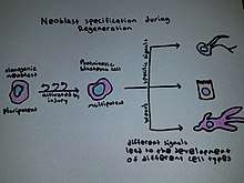
As stated above, there are several different types of organisms that can utilize a regenerative blastema as an adult. These organisms include urodele amphibians, zebrafish, and planarian flatworms as major creatures of study. In flatworms, the formation of a blastema needs adult stem cells that are called neoblasts for any type of regeneration to occur.[7] Flatworms use these undifferentiated cells for regeneration after paracrine factors can provide signals from the surface of the wound. The cells in the blastema are also referred to as clonogenic neoblasts (cNeoblasts) that are able to move to the site of the wound and reform the tissue.[8] In urodele amphibians, studies suggest that dedifferentiation of cells leads to the formation of a blastema that is able to form multiple tissue types after the amputation of their tails and wound healing occurs.[9][10] In zebrafish, and in general, it seems as if experts are still uncertain of what truly forms the blastema. However, two common theories that have often been expressed are cell dedifferentiation and the recruitment of stem cells to the wound site.[11]
Involved Signaling Pathways
There are several different signaling pathways that have been shown to be involved with limb regeneration through the formation of the blastema. In flatworms, studies suggest that after using RNA interference Smad-beta-catenin-1 was found to set up the anterior-posterior axis. Inhibitions to this results in reversed polarity across the blastema.[7] Urodeles use hedgehog for dorsal-ventral patterning of their regenerating tail and its surrounding tissue. This was suggested by its inhibition leading to reduced blastemas.[10] Zebrafish seem to use IGF signalling in limb regeneration as its inhibition led to clues of them being required for blastema function.[12]
References
- Henry George Liddell, Robert Scott, A Greek-English Lexicon, βλάστημα
- Kragl M, Knapp D, Nacu E, Khattak S, Maden M, Epperlein HH, Tanaka EM (July 2009). "Cells keep a memory of their tissue origin during axolotl limb regeneration". Nature. 460 (7251): 60–5. doi:10.1038/nature08152. PMID 19571878.
- Tanaka EM, Reddien PW (July 2011). "The cellular basis for animal regeneration". Dev. Cell. 21 (1): 172–85. doi:10.1016/j.devcel.2011.06.016. PMC 3139400. PMID 21763617.
- Godwin J (September 2014). "The promise of perfect adult tissue repair and regeneration in mammals: Learning from regenerative amphibians and fish". BioEssays. 36 (9): 861–71. doi:10.1002/bies.201300144. PMID 25043537.
- Wade, Nicholas (April 11, 2006). "Regrow Your Own". New York Times. Retrieved February 23, 2010.
- Christensen RN, Tassava RA (February 2000). "Apical epithelial cap morphology and fibronectin gene expression in regenerating axolotl limbs". Dev. Dyn. 217 (2): 216–24. doi:10.1002/(SICI)1097-0177(200002)217:2<216::AID-DVDY8>3.0.CO;2-8. PMID 10706145.
- Petersen CP, Reddien PW (January 2008). "Smed-betacatenin-1 is required for anteroposterior blastema polarity in planarian regeneration". Science. 319 (5861): 327–30. doi:10.1126/science.1149943. PMID 18063755.
- Gilbert, Scott F.; Barresi, Michael J. F. (2016). "22". Developmental Biology (11th ed.). Sunderland, Massachusetts: Sinauer Associates, Inc. pp. 701–702.
- Echeverri K, Clarke JD, Tanaka EM (August 2001). "In vivo imaging indicates muscle fiber dedifferentiation is a major contributor to the regenerating tail blastema". Dev. Biol. 236 (1): 151–64. doi:10.1006/dbio.2001.0312. PMID 11456451.
- Schnapp E, Kragl M, Rubin L, Tanaka EM (July 2005). "Hedgehog signaling controls dorsoventral patterning, blastema cell proliferation and cartilage induction during axolotl tail regeneration". Development. 132 (14): 3243–53. doi:10.1242/dev.01906. PMID 15983402.
- Nechiporuk A, Keating MT (June 2002). "A proliferation gradient between proximal and msxb-expressing distal blastema directs zebrafish fin regeneration". Development. 129 (11): 2607–17. PMID 12015289.
- Chablais F, Jazwinska A (March 2010). "IGF signaling between blastema and wound epidermis is required for fin regeneration". Development. 137 (6): 871–9. doi:10.1242/dev.043885. PMID 20179093.
Further reading
- Becker, Robert O.; Gary Selden (1998). The Body Electric. Harper Paperbacks. ISBN 0-688-06971-1.