Beta defensin
Beta defensins are a family of mammalian defensins. The beta defensins are antimicrobial peptides implicated in the resistance of epithelial surfaces to microbial colonization.
| Beta defensin | |||||||||
|---|---|---|---|---|---|---|---|---|---|
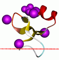 | |||||||||
| Identifiers | |||||||||
| Symbol | Defensin_beta | ||||||||
| Pfam | PF00711 | ||||||||
| InterPro | IPR001855 | ||||||||
| SCOPe | 1bnb / SUPFAM | ||||||||
| OPM superfamily | 54 | ||||||||
| OPM protein | 1ut3 | ||||||||
| |||||||||
Defensins are 2-6 kDa, cationic, microbicidal peptides active against many Gram-negative and Gram-positive bacteria, fungi, and enveloped viruses,[1] containing three pairs of intramolecular disulfide bonds. On the basis of their size and pattern of disulfide bonding, mammalian defensins are classified into alpha, beta and theta categories. Every mammalian species explored thus far has beta-defensins. In cows, as many as 13 beta-defensins exist in neutrophils. However, in other species, beta-defensins are more often produced by epithelial cells lining various organs (e.g. the epidermis, bronchial tree and genitourinary tract.
Human, rabbit and guinea-pig beta-defensins, as well as human beta-defensin-2 (hBD2), induce the activation and degranulation of mast cells, resulting in the release of histamine and prostaglandin D2.[2]
Genes
β-defensins are coding for genes which impact the function of the innate immune system.[3] These genes are responsible for production of antimicrobial peptides found in white blood cells such as macrophages, granulocytes and NK-cells, β-defensins are also found in epithelial cells.[4] Single-nucleotide polymorphisms (SNPs) are found in genes coding for β-defensins.[5] The presences of SNPs are lower in the coding regions compared to non-coding regions.[5] The appearance of SNPs in the coding region will highly likely affecting the resistance against infections through changes in the protein sequences which will give rise to different biological functions.[5]
Initiation
Receptors such as toll-like receptors (TLR) and nod-like receptors (NLR) will activate the immune system by binding of ligands such as lipopolysaccharides and peptidoglycan.[6] Toll-like receptors are expressed in intestinal epithelial cells [7] or antigen presenting cells (APCs) such as dendritic cells, B-lymphocytes and macrophages.[6] When the receptors are activated a cascade reaction will take place and substances such as cytokines and antimicrobial peptides [8] will be released.[6]
Function
β-defensins are cationic and can therefore interact with the membrane of invading microbes, which are negative due to lipopolysaccharides (LPS) and lipoteichoic acid (LTA) found in the cell membrane.[1] The peptides have higher affinity to the binding site compared to Ca2+ and Mg2+ ions.[5] The peptides will therefore exchange place with those ions, thus affecting the stability of the membrane.[5] The peptides have a greater size compared with the ions which cause changes in the membrane structure.[5] Due to changes in the electric potential, peptides will pass across the membrane and thus aggregate into dimers.[9] Pore complex will be created as a result of breaking the hydrogen bonds between the amino acids in the terminal end of the strands connecting defensins monomers.[9] Formation of pore complex will cause membrane depolarization and cell lysis.[5]
Defensins not only have the ability to strengthen the innate immune system but can also enhance the adaptive immune system by chemotaxis of monocytes, T-lymphocytes, dendritic cells and mast cells to the infection site.[5] Defensins will also improve the capacity of macrophage phagocytosis.[5]
Avian β-defensins
β-defensins are classified in three classes and Avian β-defensins constitute for one of the classes.[3] This division is based on Zhang’s classification and both the length, the homology of the peptides and the gene structure are factors affecting the classification.[9]
Avian β-defensins are separated in avian heterophiles and non-heterophiles. Avian heterophiles can be divided into two sub-classes, depending on the number of present homologous residues in the genome.[9]
Avian heterophiles lack protective oxidative mechanisms, such as superoxide and myeloperoxidase. Making non-oxidative mechanisms, such as lysosomes and cationic peptides, even more important.[9]
Evolution
Ostriches have a genome containing the gene coding for the antimicrobial peptide, Ostricacin-1. The presence of this peptide indicate that the genes coding for β-defensins have existed for a long time.[9] Ostrich and other ratite species are related to Palaeognathiformes, which is the oldest order of birds living today.[10]
β-defensins genes are found in the genome of both ostrich and mammalians.[9] The genes coding for β-defensins could originate from genes which existed prior diversification of the avian and the mammalian line, which occurred for around 150 million years ago.[11]
The fact that alpha and theta defensins are absence in older vertebrates, like birds and fishes, indicates that defensins must have evolved from the same ancestral gene coding for β-defensins.[12]
Hoover et al. (2001) showed that the origin of defensins were molecules similar to β-defensins which are found today, by comparing the amino acids and structures of the origin of β-defensins with β-defensins from insects and α-defensins found in mammalians.[13] The β-defensins found in insects were actually more similar to the origin of defensins compared to α-defensins found in mammalians. The insects lines have been around for a longer time compared to mammalian lines, which suggest that the ancestor of the genes coding for defensins have existed for a long time.[9]
The first beta-defensin discovered was Tracheal Antimicrobial Peptide, found in the bovine airway in 1991.[14] The first human beta-defensin, HBD1, was discovered in 1995,[2] followed by the HBD2 in 1997.[15]
Human proteins containing this domain
DEFB1; DEFB103A; DEFB105A; DEFB105B; DEFB106; DEFB108B; DEFB109; DEFB110; DEFB111; DEFB114; DEFB130; DEFB136; DEFB4; SPAG11A;
See also
- Defensin
- α-defensin
- β-defensin
- θ-defensin
- Lingual antimicrobial peptide
References
- White SH, Wimley WC, Selsted ME (August 1995). "Structure, function, and membrane integration of defensins". Curr. Opin. Struct. Biol. 5 (4): 521–7. doi:10.1016/0959-440X(95)80038-7. PMID 8528769.
- Bensch KW, Raida M, Mägert HJ, Schulz-Knappe P, Forssmann WG (July 1995). "hBD-1: a novel beta-defensin from human plasma". FEBS Lett. 368 (2): 331–5. doi:10.1016/0014-5793(95)00687-5. PMID 7628632.
- Hellgren O, Sheldon BC (July 2011). "Locus-specific protocol for nine different innate immune genes (antimicrobial peptides: β-defensins) across passerine bird species reveals within-species coding variation and a case of trans-species polymorphisms". Molecular Ecology Resources. 11 (4): 686–692. doi:10.1111/j.1755-0998.2011.02995.x.
- Ganz T (September 2003). "Defensins: antimicrobial peptides of innate immunity". Nat. Rev. Immunol. 3 (9): 710–20. doi:10.1038/nri1180. PMID 12949495.
- van Dijk A, Veldhuizen EJ, Haagsman HP (July 2008). "Avian defensins". Vet. Immunol. Immunopathol. 124 (1–2): 1–18. doi:10.1016/j.vetimm.2007.12.006. PMID 18313763.
- Mogensen TH (April 2009). "Pathogen recognition and inflammatory signaling in innate immune defenses". Clin. Microbiol. Rev. 22 (2): 240–73, Table of Contents. doi:10.1128/CMR.00046-08. PMC 2668232. PMID 19366914.
- Abreu MT (February 2010). "Toll-like receptor signalling in the intestinal epithelium: how bacterial recognition shapes intestinal function". Nat. Rev. Immunol. 10 (2): 131–44. doi:10.1038/nri2707. PMID 20098461.
- Vora P, Youdim A, Thomas LS, Fukata M, Tesfay SY, Lukasek K, Michelsen KS, Wada A, Hirayama T, Arditi M, Abreu MT (November 2004). "Beta-defensin-2 expression is regulated by TLR signaling in intestinal epithelial cells". J. Immunol. 173 (9): 5398–405. doi:10.404/jimmunol.173.9.5398. PMID 15494486.
- Sugiarto H, Yu PL (October 2004). "Avian antimicrobial peptides: the defense role of beta-defensins". Biochem. Biophys. Res. Commun. 323 (3): 721–7. doi:10.1016/j.bbrc.2004.08.162. PMID 15381059.
- Yu P-L, Choudhury SD, Ahrens K (January 2001). "Purification and characterization of the antimicrobial peptide, ostricacin". Biotechnology Letters. 23 (3): 207–210. doi:10.1023/A:1005623806445.
- Hedges SB, Parker PH, Sibley CG, Kumar S (May 1996). "Continental breakup and the ordinal diversification of birds and mammals". Nature. 381 (6579): 226–9. doi:10.1038/381226a0. PMID 8622763.
- Semple CA, Rolfe M, Dorin JR (2003). "Duplication and selection in the evolution of primate beta-defensin genes". Genome Biol. 4 (5): R31. doi:10.1186/gb-2003-4-5-r31. PMC 156587. PMID 12734011.
- Hoover DM, Chertov O, Lubkowski J (October 2001). "The structure of human beta-defensin-1: new insights into structural properties of beta-defensins". J. Biol. Chem. 276 (42): 39021–6. doi:10.1074/jbc.M103830200. PMID 11486002.
- Diamond, G.; Zasloff, M.; Eck, H.; Brasseur, M.; Maloy, W.; Bevins, C. (1991). "Tracheal antimicrobial peptide, a novel cysteine-rich peptide from mammalian tracheal mucosa: Peptide isolation and cloning of a cDNA". Proc. Natl. Acad. Sci. USA. 88: 3952–3956. doi:10.1073/pnas.88.9.3952. PMC 51571. PMID 2023943.
- Harder J, Siebert R, Zhang Y, Matthiesen P, Christophers E, Schlegelberger B, Schröder JM (December 1997). "Mapping of the gene encoding human beta-defensin-2 (DEFB2) to chromosome region 8p22-p23.1". Genomics. 46 (3): 472–5. doi:10.1006/geno.1997.5074. PMID 9441752.
Further reading
- Liu L, Zhao C, Heng HH, Ganz T (August 1997). "The human beta-defensin-1 and alpha-defensins are encoded by adjacent genes: two peptide families with differing disulfide topology share a common ancestry". Genomics. 43 (3): 316–20. doi:10.1006/geno.1997.4801. PMID 9268634.