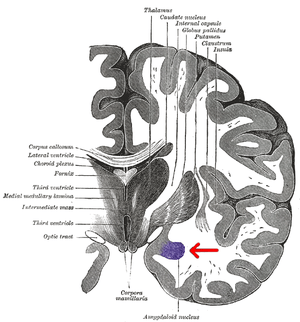
Basolateral amygdala
The basolateral amygdala (BLA) or basolateral complex, consists of the lateral, basal and accessory-basal nuclei of the amygdala. The lateral nuclei receives the majority of sensory information, which arrives directly from the temporal lobe structures, including the hippocampus and primary auditory cortex. The information is then processed by the basolateral complex and is sent as output to the central nucleus of the amygdala. This is how most emotional arousal is formed in mammals.[1]

The basolateral amygdala and nucleus accumbens shell together mediate specific Pavlovian-instrumental transfer, a phenomenon in which a classically conditioned stimulus modifies operant behavior.[2][3] The primary function of the basolateral complex is stimulating fear response. The fear system is intended to avoid pain or injury. For this reason the responses must be quick, and reflex-like. To achieve this, the “low-road” or a bottom-up process is used to generate a response to stimuli that are potentially hazardous. The stimulus reaches the thalamus, and information is passed to the lateral nucleus, then the basolateral system, and immediately to the central nucleus where a response is then formed. There is no conscious cognition involved in these responses. Other non-threatening stimuli are processed via the “high road” or a top-down form of processing.[4] In this case, the stimulus input reaches the sensory cortex first, leading to more conscious involvement in the response. In immediately threatening situations, the fight-or-flight response is reflexive, and conscious thought processing doesn’t occur until later.[5]
An important process that occurs in basolateral amygdala is consolidation of cued fear memory. One proposed molecular mechanism for this process is collaboration of M1-Muscarinic receptors, D5 receptors and beta-2 adrenergic receptors to redundantly activate phospholipase C, which inhibits the activity of KCNQ channels[6] that conduct inhibitory M current.[7] The neuron then becomes more excitable and the consolidation of memory is enhanced.[6]
The amygdala has several different nuclei and internal pathways; the basolateral complex (or basolateral amygdala), the central nucleus, and the cortical nucleus are the most well-known. Each of these has a unique function and purpose within the amygdala.
References
- Baars, B. J, Gage, N. M. (2010). Cognition, Brain, and Consciousness: introduction to cognitive neuroscience second edition. Academic Press. Burlington MA
- Cartoni E, Puglisi-Allegra S, Baldassarre G (November 2013). "The three principles of action: a Pavlovian-instrumental transfer hypothesis". Frontiers in Behavioral Neuroscience. 7: 153. doi:10.3389/fnbeh.2013.00153. PMC 3832805. PMID 24312025.
- Salamone JD, Pardo M, Yohn SE, López-Cruz L, SanMiguel N, Correa M (2016). "Mesolimbic Dopamine and the Regulation of Motivated Behavior". Current Topics in Behavioral Neurosciences. 27: 231–257. doi:10.1007/7854_2015_383. PMID 26323245.
Considerable evidence indicates that accumbens DA is important for Pavlovian approach and Pavlovian-to-instrumental transfer [(PIT)] ... PIT is a behavioral process that reflects the impact of Pavlovian-conditioned stimuli (CS) on instrumental responding. For example, presentation of a Pavlovian CS paired with food can increase output of food-reinforced instrumental behaviors, such as lever pressing. Outcome-specific PIT occurs when the Pavlovian unconditioned stimulus (US) and the instrumental reinforcer are the same stimulus, whereas general PIT is said to occur when the Pavlovian US and the reinforcer are different. ... More recent evidence indicates that accumbens core and shell appear to mediate different aspects of PIT; shell lesions and inactivation reduced outcome-specific PIT, while core lesions and inactivation suppressed general PIT (Corbit and Balleine 2011). These core versus shell differences are likely due to the different anatomical inputs and pallidal outputs associated with these accumbens subregions (Root et al. 2015). These results led Corbit and Balleine (2011) to suggest that accumbens core mediates the general excitatory effects of reward-related cues. PIT provides a fundamental behavioral process by which conditioned stimuli can exert activating effects upon instrumental responding
- Breedlove, S., Watson, N. (2013). Biological Psychology: an introduction to behavioral cognitive, and clinical neuroscience Seventh edition. Sinauer Associates, inc. Sunderland: MA.
- Smith, C., & Kirby, L., (2001). Toward delivering on the promise of appraisal theory. In K. R. Scherer, a. Schorr, & T. Johnstone (Eds.), Apraisal processes in emotion : Theory, methods, research. Oxford, UK: Oxford University Press.
- Young, M. B.; Thomas, S. A. (2014). "M1-muscarinic receptors promote fear memory consolidation via phospholipase C and the M-current". Journal of Neuroscience. 34 (5): 1570–8. doi:10.1523/JNEUROSCI.1040-13.2014. PMC 3905134. PMID 24478341.
- Schroeder, B. C.; Hechenberger, M; Weinreich, F; Kubisch, C; Jentsch, T. J. (2000). "KCNQ5, a novel potassium channel broadly expressed in brain, mediates M-type currents". Journal of Biological Chemistry. 275 (31): 24089–95. doi:10.1074/jbc.M003245200. PMID 10816588.