Aortic valve replacement
Aortic valve replacement is a procedure whereby the failing aortic valve of a patient's heart is replaced with an artificial heart valve. The aortic valve may need to be replaced because:
- The valve is leaky (aortic insufficiency, also known as aortic regurgitation)
- The valve is narrowed and doesn't open fully (aortic stenosis)
| Aortic valve replacement | |
|---|---|
| ICD-9-CM | 35.21-35.22V43.3 |
Current methods for aortic valve replacement include open heart surgery, minimally invasive cardiac surgery (MICS) and transcatheter aortic valve replacement (TAVR).
History
During the late 1940s and early 1950s, the first surgical approaches towards treating aortic valve stenosis had limited success. The first attempts were valvotomies, (i.e. cutting the valve while the heart is pumping). A ball valve prosthesis placed on the descending thoracic aorta (heterotopically) was developed by Hufnagel, Harvey and others to address aortic stenosis, but had disastrous complications. Later, with the innovation of cardiopulmonary bypass, the ball valve prosthesis was placed orthotopically (i.e. in same place as the original aortic valve). This first generation of prosthetic valves was durable, but needed intense anti-coagulation, and cardiac hemodynamics were compromised. During the mid-1950s, a single-leaflet prosthesis was developed by Bahnson et al. In early 1960, Ross and Barratt-Boyes used allografts. Tissue prosthetic valves were introduced in 1965 by Binet in Paris, but they degenerated quickly because the tissue was insufficiently preserved. Carpentier solved this problem by introducing glutaraldehyde-preserved stent-mounted porcine valves.[1][2]
Anatomy, physiology and pathophysiology
| U/S findings | Mild | Moderate | Severe |
|---|---|---|---|
| Aortic valve area, cm2 | >1.5 | 1.0-1.5 | <1.0 |
| Aortic valve area index, cm2/m2 | - | - | 0.6 |
| Mean pressure gradient, mmHg | 25 | 25-40 | >40 |
| Peak jet velocity, m/s | <3 | 3-4 | >4 |
| U/S findings | Mild | Moderate | Severe |
|---|---|---|---|
| Jet width of left ventricular outflow tract | <25% | 25–65% | >65% |
| Vena contracta width, cm | <0.3 | 0.3–0.6 | 0.6 |
| Regurgitant volume, mL per beat | <30 | 30–49 | 60 |
| Regurgitant fraction, % | 30 | 30–49 | >50 |
| Regurgitant orifice area, cm2 | <0.10 | 0.10–0.30 | >0.30 |
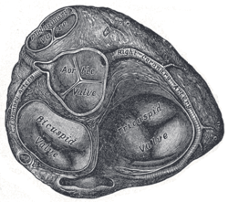
The aortic valve is semilunar (half-moon shaped) with three cusps. It separates the heart from the aorta. Each cusp is attached to the aortic wall creating a sinus; a Valsalva sinus. The origins of the two coronary arteries are sited in two Valsalva sinuses, each named after the coronary artery they supply. Leaflets are separated by commissures. The posterior leaflet is in continuation with the anterior leaflet of the mitral valve (the tissue is called the aorto-mitral curtain).[3] The aortic valve is opened during systole, the driving force for it to open is the difference in pressure between the contracting left ventricle of the heart and the aorta. During cardiac diastole (when the heart chamber gets bigger) the aortic valve closes.[4]
Aortic stenosis most commonly is the result of calcification of the cusps. Other reasons for stenosis are the bicuspid valve (some patients have only two cusps at the aortic valve instead of the usual three) and rheumatic aortic stenosis (now rare in the West). Obstruction at the level of the aortic valve causes increased pressure within the heart's left ventricle. This can lead to hypertrophy and ultimately dysfunction of the heart. While x-ray and ECG might indicate aortic stenosis, echocardiography is the diagnostic procedure of choice. US findings also help in grading the severity of the disease. In cases of symptomatic severe aortic stenosis, AVR is warranted. In cases of asymptomatic but severe aortic stenosis, more factors should be taken into consideration.[5]
Aortic regurgitation, on the other hand, has many causes: degeneration of the cusps, endocarditis, bicuspid aortic valve, aortic root dilatation, trauma, connective tissue disorders such as Marfan syndrome or Ehlers-Danlos lead to imperfect closure of the valve during diastole, hence the blood is returning from the aorta towards the left ventricle of the heart. Acute aortic regurgitation (caused by endocarditis, aortic dissection or trauma) ends up in pulmonary edema, because of the acute increase in left ventricle (LVEDP) that does not have time to adjust to the regurgitation. Chronic regurgitation, by contrast, gives the heart time to change shape, resulting in eccentric hypertrophy, which has disastrous effects on the myocardium. Ultrasound is here also the best diagnostic mobility, either it is transthoracic or transesophageal. [6]
Indications for surgery
Guidelines for aortic valve replacement
As long-term data on the survival and quality of life of people following valve replacement have become available, evidence-based guidelines for aortic valve replacement have been developed. These help healthcare professionals decide when aortic valve replacement is the best option for a patient. Two widely accepted sets of guidelines used by surgeons and cardiologists are the American Heart Association and American College of Cardiology Guidelines for the Management of Patients with Valvular Heart Disease,[7] and the European Society of Cardiology and the European Association for Cardio‑Thoracic Surgery Guidelines for the management of valvular heart disease.[8]
Aortic stenosis
Aortic stenosis is treated with aortic valve replacement in order to avoid angina, syncope, or congestive heart failure. Individuals with severe aortic stenosis are candidates for aortic valve replacement once they develop symptoms or when their heart function is impacted. Some people with asymptomatic aortic stenosis may also be candidates for aortic valve replacement, especially if symptoms appear during exercise testing.[8] Patients with moderate aortic valve stenosis who need another type of cardiac surgery (i.e. coronary artery bypass surgery) should also have their valve addressed by the surgical team if echocardiography unveils significant heart problems.[9]
Low gradient aortic stenosis with concomitant left ventricular dysfunction poses a significant question to the surgeon and the patient. Stress echocardiography (i.e. with dobutamine infusion) can help determine if the ventricle is dysfunctional because of aortic stenosis, or because the myocardium lost its ability to contract.[10]
Aortic insufficiency
Many people with aortic insufficiency often don’t develop symptoms until they have had the condition for many years.[11] Aortic valve replacement is indicated for symptoms such as shortness of breath, and in cases where the heart has begun to enlarge (dilate) from pumping the increased volume of blood that leaks back through the valve.[8]
Types of valves
There are two basic types of replacement heart valve: tissue (bioprosthetic) valves and mechanical valves.[12]
Tissue valves
Tissue heart valves are usually made from animal tissue (heterografts) mounted on a metal or polymer support.[13] Bovine (cow) tissue is most commonly used, but some are made from porcine (pig) tissue.[14] The tissue is treated to prevent rejection and calcification (where calcium builds up on the replacement valve and stops it working properly).[15]
Occasionally, alternatives to animal tissue valves are used: aortic homografts and pulmonary autografts. An aortic homograft is an aortic valve from a human donor, retrieved either after their death or from their heart if they are undergoing a heart transplant.[16] A pulmonary autograft, also known as the Ross procedure is where the aortic valve is removed and replaced with the patient's own pulmonary valve (the valve between the right ventricle and the pulmonary artery). A pulmonary homograft (a pulmonary valve taken from a cadaver) is then used to replace the patient's own pulmonary valve. This procedure was first performed in 1967 and is used primarily in children, as it allows the patient's own pulmonary valve (now in the aortic position) to grow with the child.[16]
Tissue valves can last 10–20 years.[17] However, they tend to deteriorate more quickly in younger patients.[18] New ways of preserving the tissue for longer are being investigated. One such preservation treatment is now being used in a commercially available tissue heart valve. In sheep and rabbit studies, the tissue (called RESILIA™ tissue) had less calcification than control tissue.[29][30] However, long-term durability data in patients are not yet available.[31]
Tissue valves come as stented or stentless. Stented valves come in sizes from 19 mm to 29 mm.[19] Stentless valves are directly sutured at the aortic root. The major advantage of stentless valves is that they limit patient–prosthesis mismatch (when the area of the prosthetic valve is too small in relation to the size of the patient, increasing the pressure inside the valve[20]) and can be helpful when dealing with small aortic root. Their disadvantage is that it is more time-consuming to implant stentless valves than stented valves.[21]
Mechanical valves
Mechanical valves are made from synthetic materials, such as titanium or pyrolytic carbon.[22] They are more durable than tissue valves, typically lasting 20–30 years.[12] However, the risk of blood clots forming is higher with mechanical valves than with tissue valves. As a result, people with mechanical heart valves must take anticoagulant (blood-thinning) drugs, such as warfarin, for the rest of their lives, making them more prone to bleeding.[12] The sound of the valve can sometimes be heard (often as clicks) and can be disturbing.[23]
Valve selection
Valve choice is a balance between the lower durability of tissue valves and the increased risk of blood clots and bleeding with mechanical valves. Guidelines suggest that patient age, lifestyle and medical history should all be considered when choosing a valve. Tissue valves deteriorate more rapidly in young patients and during pregnancy, but they are preferable for women who wish to have children because pregnancy increases the risk of blood clots. Typically, a mechanical valve is considered for patients under 60 years old, while a tissue valve is considered for patients over the age of 65 years.[12]
Procedure
Surgical Procedure
.svg.png)
Aortic valve replacement is conventionally done through a median sternotomy, meaning the incision is made by cutting through the breastbone (sternum). Once the protective membrane around the heart (pericardium) has been opened, the patient is cannulated (aortic cannulation by a cannula placed on the aorta and a venous canulation by a single atrial venous cannula inserted through the right atrium. The patient is put on a cardiopulmonary bypass machine, also known as the heart–lung machine. This machine breathes for the patient and pumps their blood around their body while the surgeon replaces the heart valve.
Once on cardiopulmonary bypass, the patient's heart is stopped (cardioplegia). This can be done with a Y-type cardioplegic infusion catheter placed on the aorta, de-aired and connected to the cardiopulmonary bypass machine. Alternatively, a retrograde cardioplegic cannula can be inserted at the coronary sinus. Some surgeons also opt to place a vent in the left ventricle through the right superior pulmonary vein, because this helps to prevent left ventricular distention before and after cardiac arrest. When the set-up is ready, the aorta is clamped shut with a cross-clamp to stop blood pumping through the heart and cardioplegia is infused. The surgeon incises the aorta a few milometers above the sinotubular junction (just above the coronary ostia, where the coronary arteries join the aorta) – a process known as aortotomy. After this, cardioplegia is delivered directly through the ostia.[24][25]
The heart is now still and the surgeon removes the patient's diseased aortic valve. The cusps of the aortic valve are excised, and calcium is removed (debrided) from the aortic annulus. The surgeon measures the size of the aortic annulus and fits a mechanical or tissue valve of the appropriate size. Usually the valve is fixed in place with sutures, although some sutureless valves are available. If the patient's aortic root is very small, the sutures are placed outside of the aortic root instead of at the annulus, to gain some extra space.
Once the valve is in place and the aorta has been closed, patient is placed in a Trendelenburg position and the heart is de-aired and restarted. The patient is taken off the cardiopulmonary bypass machine. Transesophageal echocardiogram (an ultrasound of the heart done through the esophagus) can be used to verify that the new valve is functioning properly. Pacing wires are usually put in place, so that the heart can be manually controlled should any complications arise after surgery. Drainage tubes are also inserted, to drain fluids from the chest. These are usually removed within 36 hours, while the pacing wires are generally left in place until right before the patient is discharged from the hospital.[24][25]
Hospital stay and recovery time
After aortic valve replacement, the patient will usually stay in an intensive care unit for 12–36 hours. Unless complications arise, the patient is then able to go home after approximately four to seven days.[26] Common complications include disturbances to the heart’s rhythm (heart block), which typically require the permanent insertion of a cardiac pacemaker.[27]
Recovery from aortic valve replacement takes about three months if the patient is in good health. Patients are advised not to lift anything heavier than 10 lbs for several weeks, and not to do any heavy lifting for 4–6 months after surgery to avoid damaging their breastbone. Often patients will be referred to participate in cardiopulmonary rehabilitation, which optimizes recovery and physical function in patients with recent cardiac surgeries. This can be done in an outpatient setting.[28]
Outcomes
Surgery usually relieves the aortic disease symptoms that led the patient to the operating room. The survival curve of patients that undergo aortic valve replacements is slightly inferior to the curve of their corresponding healthy same-aged same sex population.[29] (Pre-operative) severe left ventricular hypertrophy is a contributing factor to morbidity.[29]
The risk of dying as a result of aortic valve replacement is estimated at 1–3%.[30][31][32] Combining aortic valve replacement with coronary artery bypass grafting increases the risk of mortality.[30] Older patients, as well as those who are frail and/or have other health problems (comorbidities), have a higher risk of experiencing complications.[31] Possible problems include cardiac infarction/failure, arrhythmia or heart block (typically requires the permanent insertion of a cardiac pacemaker), mediastinal bleeding, stroke and infection. Late complications include endocarditis, thromboembolic events (blood clots), prosthetic valve dysfunction and paravalvular leak (blood flowing between the edge of the prosthetic valve and the cardiac tissue).[29]
Patient–prosthesis mismatch
When dealing with a small aortic annulus, the surgeon might be forced to insert a small prosthetic aortic valve, with an orifice that is too small in relation to the size of the patient. This is known as patient–prosthesis mismatch. It increases the pressure of the blood flowing through the valve and can negatively affect outcomes.[20] Various techniques and stentless valves have been utilized to avoid this phenomenon.[33]
Less invasive procedures
Minimally invasive cardiac surgery
Since the late 1990s, some cardiac surgeons have been performing aortic valve replacement procedures using an approach referred to as minimally invasive cardiac surgery (MICS).[34] Using this approach, the surgeon replaces the valve through a smaller chest incision (6–10 cm) than that for a median sternotomy. MICS typically involves shorter recovery times and more attractive cosmetic results.[35]
Transcatheter aortic valve replacement
Another alternative for many high-risk or elderly patients is transcatheter aortic valve replacement (TAVR, also known as TAVI, transcatheter aortic valve implantation). Rather than removing the existing valve, this technique pushes a new valve into the place of the existing valve. The replacement valve is collapsible and is delivered to the site of the existing valve through a tube called a catheter. The catheter may be inserted through the femoral artery in the thigh (transfemoral approach), or using a small incision in the chest and then through a large artery or the tip of the left ventricle (transapical approach).[36] Fluoroscopy and TTE are utilized for the positioning of the valve.[36] Once the replacement valve is in place, it is expanded, pushing the old valve’s leaflets out of the way.[37]
Guidelines suggest TAVR for most patients aged 75 years and older, and surgical aortic valve replacement for most patients aged less than 75 years.[38] Ultimately, the best treatment choice is a decision based on many individual factors.[39][38]
See also
References
- Kouchoukos 2012, p. 543.
- Emery 2017, pp. 649-652.
- Brzezinski 2017, p. 663.
- Brzezinski 2017, p. 636.
- Brzezinski 2017, pp. 636-637.
- Brzezinski 2017, pp. 638-643.
- Nishimura, Rick A.; Otto, Catherine M.; Bonow, Robert O.; Carabello, Blase A.; Erwin, John P.; Fleisher, Lee A.; Jneid, Hani; Mack, Michael J.; McLeod, Christopher J. (2017). "2017 AHA/ACC Focused Update of the 2014 AHA/ACC Guideline for the Management of Patients With Valvular Heart Disease". Journal of the American College of Cardiology. 70 (2): 252–289. doi:10.1016/j.jacc.2017.03.011. PMID 28315732.
- Baumgartner, Helmut; Falk, Volkmar; Bax, Jeroen J; De Bonis, Michele; Hamm, Christian; Holm, Per Johan; Iung, Bernard; Lancellotti, Patrizio; Lansac, Emmanuel (2017-09-21). "2017 ESC/EACTS Guidelines for the management of valvular heart disease". European Heart Journal. 38 (36): 2739–2791. doi:10.1093/eurheartj/ehx391. ISSN 0195-668X. PMID 28886619.
- Yanagawa 2017, pp. 665-668.
- Fullerton 2014, p. 463.
- Maurer, G. (2006-05-02). "Aortic regurgitation". Heart. 92 (7): 994–1000. doi:10.1136/hrt.2004.042614. ISSN 1355-6037. PMC 1860728. PMID 16775114.
- Tillquist; Tillquist; Maddox, Tom (2011). "Cardiac crossroads: deciding between mechanical or bioprosthetic heart valve replacement". Patient Preference and Adherence. 5: 91–9. doi:10.2147/PPA.S16420. ISSN 1177-889X. PMC 3063655. PMID 21448466.
- Pibarot, Philippe; Dumesnil, Jean G. (2009-02-24). "Prosthetic Heart Valves: Selection of the Optimal Prosthesis and Long-Term Management". Circulation. 119 (7): 1034–1048. doi:10.1161/CIRCULATIONAHA.108.778886. ISSN 0009-7322. PMID 19237674.
- Hickey, Graeme L.; Grant, Stuart W.; Bridgewater, Ben; Kendall, Simon; Bryan, Alan J.; Kuo, James; Dunning, Joel (2015). "A comparison of outcomes between bovine pericardial and porcine valves in 38 040 patients in England and Wales over 10 years". European Journal of Cardio-Thoracic Surgery. 47 (6): 1067–1074. doi:10.1093/ejcts/ezu307. ISSN 1873-734X. PMID 25189704.
- Li, Kan Yan Chloe (2019-04-11). "Bioprosthetic Heart Valves: Upgrading a 50-Year Old Technology". Frontiers in Cardiovascular Medicine. 6: 47. doi:10.3389/fcvm.2019.00047. ISSN 2297-055X. PMC 6470412. PMID 31032263.
- Bloomfield, P. (2002-06-01). "Choice of heart valve prosthesis". Heart. 87 (6): 583–589. doi:10.1136/heart.87.6.583. PMC 1767148. PMID 12010950.
- Harris, Christopher; Croce, Beth; Cao, Christopher (2015-10-07). "Tissue and mechanical heart valves". Annals of Cardiothoracic Surgery. 4 (4): 399. doi:10.3978/6884 (inactive 2019-11-24). ISSN 2225-319X. PMC 4526499. PMID 26309855.
- Johnston, Douglas R.; Soltesz, Edward G.; Vakil, Nakul; Rajeswaran, Jeevanantham; Roselli, Eric E.; Sabik, Joseph F.; Smedira, Nicholas G.; Svensson, Lars G.; Lytle, Bruce W. (2015). "Long-Term Durability of Bioprosthetic Aortic Valves: Implications From 12,569 Implants". The Annals of Thoracic Surgery. 99 (4): 1239–1247. doi:10.1016/j.athoracsur.2014.10.070. PMC 5132179. PMID 25662439.
- Sabiston 2010, p. 1189.
- Pibarot, P (2006-08-01). "Prosthesis-patient mismatch: definition, clinical impact, and prevention". Heart. 92 (8): 1022–1029. doi:10.1136/hrt.2005.067363. ISSN 1355-6037. PMC 1861088. PMID 16251232.
- Sabiston 2010, p. 1191.
- Gott, Vincent L; Alejo, Diane E; Cameron, Duke E (December 2003). "Mechanical heart valves: 50 years of evolution". The Annals of Thoracic Surgery. 76 (6): S2230–S2239. doi:10.1016/j.athoracsur.2003.09.002. ISSN 0003-4975. PMID 14667692.
- Golczyk, Karl Jan. (2010). Heart valve sound of various mechanical composite grafts, and the impact on patients' quality of life. [Verlag nicht ermittelbar]. OCLC 742549155.
- Kouchoukos 2012, p. 554.
- Emery 2017, pp. 652-653.
- Kouchoukos 2012, p. 190 & 590.
- Kim, You Ho; Lee, Jae Won; Chung, Cheol Hyun; Choi, Kee Joon; Nam, Gi Byoung; Choo, Suk Jung; Jung, Sung-Ho; Kim, Joon Bum; Hwang, Jongmin (2017-11-01). "Conduction disturbance after isolated surgical aortic valve replacement in degenerative aortic stenosis". The Journal of Thoracic and Cardiovascular Surgery. 154 (5): 1556–1565.e1. doi:10.1016/j.jtcvs.2017.05.101. ISSN 0022-5223. PMID 28712585.
- "What is Cardiac Rehabilitation?". www.heart.org. Retrieved 2019-07-30.
- Sabiston 2010, p. 1204.
- Shahian, David M.; Wormuth, David W.; Paone, Gaetano; Fernandez, Felix G.; Badhwar, Vinay; Jacobs, Jeffrey P.; D’Agostino, Richard S. (2019-01-01). "The Society of Thoracic Surgeons Adult Cardiac Surgery Database: 2019 Update on Outcomes and Quality". The Annals of Thoracic Surgery. 107 (1): 24–32. doi:10.1016/j.athoracsur.2018.10.004. ISSN 0003-4975. PMID 30423335.
- "Aortic valve replacement - Risks". nhs.uk. 2017-10-20. Retrieved 2019-07-30.
- Fullerton 2014, p. 461.
- Fullerton 2014, p. 465.
- Merk, Denis R.; Lehmann, Sven; Holzhey, David M.; Dohmen, Pascal; Candolfi, Pascal; Misfeld, Martin; Mohr, Friedrich W.; Borger, Michael A. (2015). "Minimal invasive aortic valve replacement surgery is associated with improved survival: a propensity-matched comparison†". European Journal of Cardio-Thoracic Surgery. 47 (1): 11–17. doi:10.1093/ejcts/ezu068. ISSN 1873-734X. PMID 24599160.
- Glauber, Mattia; Ferrarini, Matteo; Miceli, Antonio (2015-01-16). "Minimally invasive aortic valve surgery: state of the art and future directions". Annals of Cardiothoracic Surgery. 4 (1): 26–32–32. doi:10.3978/5476 (inactive 2019-11-24). ISSN 2225-319X. PMC 4311160. PMID 25694973.
- Sabiston 2010, p. 1203.
- "What is TAVR?". www.heart.org. Retrieved 2019-07-30.
- Spencer, Frederick A.; Mertz, Ray; Shapiro, Michael; Foroutan, Farid; Price, Susanna; Aertgeerts, Bert; Brieger, David; Vartdal, Trond; Whitlock, Richard (2016-09-28). "Transcatheter or surgical aortic valve replacement for patients with severe, symptomatic, aortic stenosis at low to intermediate surgical risk: a clinical practice guideline". BMJ. 354: i5085. doi:10.1136/bmj.i5085. ISSN 1756-1833. PMID 27680583.
- Lytvyn, Lyubov; Guyatt, Gordon H; Manja, Veena; Siemieniuk, Reed A; Zhang, Yuan; Agoritsas, Thomas; Vandvik, Per O (2016). "Patient values and preferences on transcatheter or surgical aortic valve replacement therapy for aortic stenosis: a systematic review". BMJ Open. 6 (9): e014327. doi:10.1136/bmjopen-2016-014327. ISSN 2044-6055. PMC 5051506. PMID 27687903.
Sources
- Brzezinski, Anna (28 August 2017). "Pathophysiology of Aortic Valve Disease". In Lawrence H. Cohn; David H. Adams (eds.). Cardiac Surgery in the Adult Fifth Edition. McGraw-Hill Education. ISBN 978-0-07-184487-1.
- Emery, Robert W. (28 August 2017). "Aortic Valve Replacement with a Mechanical Cardiac Valve Prosthesis". In Lawrence H. Cohn; David H. Adams (eds.). Cardiac Surgery in the Adult Fifth Edition. McGraw-Hill Education. ISBN 978-0-07-184487-1.
- Fullerton, David A (2014). "Aortic Valve Replacement". In Larry Kaiser; Irving L. Kron; Thomas L. Spray (eds.). Mastery of Cardiothoracic Surgery. Lippincott Williams & Wilkins. ISBN 978-1-4511-1315-0.
- Gao, Changqing (2014). Robotic Cardiac Surgery. Springer Netherlands. ISBN 978-94-007-7659-3.
- Kouchoukos, Nicholas T. (27 September 2012). Kirklin/Barratt-Boyes Cardiac Surgery E-Book. Elsevier Health Sciences. ISBN 978-1-4557-4605-7.
- Pibarot, Philippe; Dumesnil, Jean G. (2009-02-24). "Prosthetic Heart Valves". Circulation. Ovid Technologies (Wolters Kluwer Health). 119 (7): 1034–1048. doi:10.1161/circulationaha.108.778886. ISSN 0009-7322. PMID 19237674.
- Sabiston (16 December 2010). Sabiston and Spencer's Surgery of the Chest E-Book. Elsevier Health Sciences. ISBN 978-1-4557-0009-7.
- Yanagawa, Bobby (28 August 2017). "Stented Bioprosthetic Aortic Valve Replacement". In Lawrence H. Cohn; David H. Adams (eds.). Cardiac Surgery in the Adult Fifth Edition. McGraw-Hill Education. ISBN 978-0-07-184487-1.