Aortic hiatus
The aortic hiatus is a hole in the diaphragm. It is the lowest and most posterior of the large apertures.
| Aortic hiatus | |
|---|---|
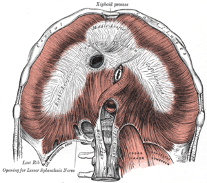 The diaphragm. Under surface. (Aortic hiatus labeled near center.) | |
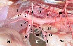 Celiac and cranial mesenteric ganglion in a cat. 1 Crus sinistrum (Diaphragma), 2 hiatus aorticus, 3 Aorta, 4 Arteria lumbalis, 5 Nervus splanchnicus major, 6 Arteria coeliaca, 7 Arteria phrenica caudalis, 8 Ganglion coeliacum, 9 Plexus coeliacus, 10 Ganglion mesentericum craniale, 11 Plexus mesentericus cranialis, 12 Arteria mesenterica cranialis, 13 Nervus splanchnicus minor, 14 Adrenal gland, 15 Arteria abdominalis cranialis, 16 Stomach, 17 Liver (Lobus caudatus), 18 Kidney | |
| Details | |
| Identifiers | |
| Latin | hiatus aorticus |
| TA | A04.4.02.010 |
| FMA | 58288 |
| Anatomical terminology | |
It is located approximately at the level of the twelfth thoracic vertebra (T12).
Structure
Strictly speaking, it is not an aperture in the diaphragm but an osseoaponeurotic opening between it and the vertebral column, and therefore behind the diaphragm (Meaning that diaphragmatic contractions do not directly influence the Aorta or aortic supply).
Occasionally some tendinous fibers prolonged across the bodies of the vertebræ from the medial parts of the inferior ends of the crura pass posterior to the aorta, and thus convert the hiatus into a fibrous ring.
The hiatus is situated slightly to the left of the mid line, and is bound anteriorly by the crura, and posteriorly by the body of the first lumbar vertebra.
Structures passing through
Through it passes the aorta and the thoracic duct
The azygos vein and hemi-azygos vein passes through the medial lumbar gapcrus of diaphragm.
Additional images
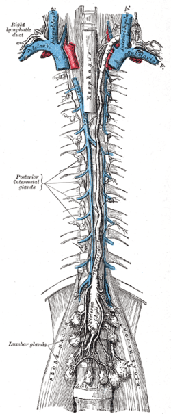 The thoracic and right lymphatic ducts.
External links
- Anatomy photo:40:08-0103 at the SUNY Downstate Medical Center - "Major Openings in the Diaphragm"
- Anatomy image:8906 at the SUNY Downstate Medical Center
| Authority control |
|---|