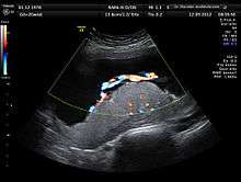
Anomaly scan
The anomaly scan, also sometimes called the anatomy scan, 20 week ultrasound, or level 2 ultrasound, is a pregnancy ultrasound performed between 18–22 weeks of gestational age. The International Society of Ultrasound in Obstetrics and Gynecology (ISUOG) recommends that this ultrasound is performed as a matter of routine prenatal care. The function of the ultrasound is to measure the fetus so that growth abnormalities can be recognized quickly later in pregnancy, and to assess for congenital malformations and multiple pregnancies (i.e. twins).[1]
| Anomaly scan | |
|---|---|
| Medical diagnostics | |
| Synonyms | 20 week ultrasound |
| Purpose | pregnancy scan for congenital malformation |
Measures assessed
The anomaly scan allows the developing fetus to be observed in terms of morphology, bone shape, skeletal features, fetal heart function, volume evaluation, fetal lung maturity[2] and general fetal well being.[3]
A standard anatomy scan typically includes
- Fetal number, including number of amnionic sacs and chorionic sacs for multiple gestations
- Fetal cardiac activity
- Fetal position relative to the uterus and cervix
- Location and appearance of the placenta, including site of umbilical cord insertion when possible
- Amnionic fluid volume
- Gestational age assessment
- Fetal weight estimation
- Fetal anatomical survey
- Evaluation of the maternal uterus, tubes, ovaries, and surrounding structures when appropriate[4]
Anomalies looked for in the fetal anatomical survey
Second-trimester ultrasound screening for aneuploidies, such as Edwards syndrome and Patau syndrome, are based on looking for soft markers and some predefined structural abnormalities. Soft markers are variations from normal anatomy, which are more common in aneuploid fetuses compared to euploid ones. These markers are often not clinically significant and do not cause adverse pregnancy outcomes.[5]
The anomalies typically looked for in an anomaly scan include:
Sex determination
Whilst sex can technically be determined earlier, sex determination at this scan is generally reliable, and so this is commonly when parents can learn the sex of their baby.
See also
References
- "Practice guidelines for performance of the routine mid-trimester fetal ultrasound scan" (PDF). ISUOG.org. Retrieved December 4, 2017.
- Bhanu Prakash, K.N.; Ramakrishnan, A.G.; Suresh, S.; Chow, T.W.P. (March 2002). "Fetal lung maturity analysis using ultrasound image features" (PDF). IEEE Transactions on Information Technology in Biomedicine. 6 (1): 38–45. doi:10.1109/4233.992160. PMID 11936595.
- Layyous, Najeeb. "The Clinical Advantages of 3D and 4D Ultrasound". www.layyous.com. Retrieved 2018-08-23.
- Cunningham, F; Leveno, KJ; Bloom, SL; Spong, CY; Dashe, JS; Hoffman, BL; Casey BM, BM; Sheffield, JS (2013). "Fetal Imaging". Williams Obstetrics, Twenty-Fourth Edition. McGraw-Hill.
- Zare Mehrjardi, Mohammad; Keshavarz, Elham (2017-04-16). "Prefrontal Space Ratio—A Novel Ultrasound Marker in the Second Trimester Screening for Trisomy 21: Systematic Review and Meta-Analysis". Journal of Diagnostic Medical Sonography. 33 (4): 269–277. doi:10.1177/8756479317702619.
- NHS Choices. "Mid-pregnancy anomaly scan - Pregnancy and baby - NHS Choices". www.nhs.uk. Retrieved 2017-12-04.