Angular gyrus
The angular gyrus is a region of the brain lying mainly in the anterolateral region of parietal lobe, that lies near the superior edge of the temporal lobe, and immediately posterior to the supramarginal gyrus. Its significance is in transferring visual information to Wernicke's area, in order to make meaning out of visually perceived words.[1] It is also involved in a number of processes related to language, number processing and spatial cognition, memory retrieval, attention, and theory of mind. It is Brodmann area 39 of the human brain.
| Angular gyrus | |
|---|---|
 | |
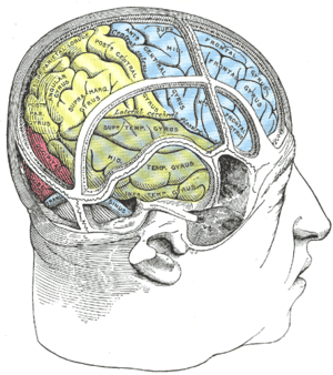 Drawing of a cast to illustrate the relations of the brain to the skull. (Angular gyrus labeled at upper left, in yellow section.) | |
| Details | |
| Identifiers | |
| Latin | gyrus angularis |
| NeuroNames | 109 |
| NeuroLex ID | birnlex_1376 |
| TA | A14.1.09.124 |
| FMA | 61898 |
| Anatomical terms of neuroanatomy | |
Anatomy
Left and right angular gyri are connected by the dorsal splenium and isthmus of the corpus callosum.[2] Both gyri lie between the four lobes.
| Connected To The | Via the |
|---|---|
| ipsilateral frontal and caudallateral prefrontal and inferior frontal regions | superior longitudinal fasciculus.[3] |
| caudate | inferior occipitofrontal fasciculus[4] |
| parahippocampal gyrus[5] and hippocampus[4] | inferior longitudinal fasciculus |
| precuneus and superior frontal gyrus | occipitofrontal fasciculus,[6] |
| supramarginal gyrus | local arcuate[7] |
Function
The angular gyrus is the part of the brain associated with complex language functions (i.e. reading, writing and interpretation of what is written). Lesion to this part of the brain shows symptoms of the Gerstmann syndrome: effects include finger agnosia, alexia (inability to read), acalculia (inability to use arithmetic operations), agraphia (inability to copy), and left-right confusion.
Language
Geschwind proposed that written word is translated to internal monologue via the angular gyrus.
V. S. Ramachandran, and Edward Hubbard published a paper in 2003 in which they speculated that the angular gyrus is at least partially responsible for understanding metaphors. They stated:
There may be neurological disorders that disturb metaphor and synaesthesia. This has not been studied in detail but we have seen disturbances in the Bouba/Kiki effect (Ramachandran & Hubbard, 2001a) as well as with proverbs in patients with angular gyrus lesions. It would be interesting to see whether they have deficits in other types of synaesthetic metaphor, e.g. 'sharp cheese' or 'loud shirt'. There are also hints that patients with right hemisphere lesions show problems with metaphor. It is possible that their deficits are mainly with spatial metaphors, such as 'He stepped down as director'.[8]
The fact that the angular gyrus is proportionately much larger in hominids than other primates, and its strategic location at the crossroads of areas specialized for processing touch, hearing and vision, leads Ramachandran to believe that it is critical both to conceptual metaphors and to cross-modal abstractions more generally. However, recent research challenges this theory.
Research by Krish Sathian (Emory University) using functional magnetic resonance imaging (fMRI) suggests that the angular gyrus does not play a role in creating conceptual metaphors. Sathian theorizes that conceptual metaphors activate the texture-selective somatosensory cortex in the parietal operculum.[9]
Brownsett and Wise highlight the role of the left angular gyrus in both speaking and writing.[10]
Mathematics and spatial cognition
Since 1919, brain injuries to the angular gyrus have been known to often cause arithmetic deficits.[11][12] Functional imaging has shown that while other parts of the parietal lobe bilaterally are involved in approximate calculations due to its link with spatiovisual abilities, the left angular gyrus together with left Inferior frontal gyrus are involved in exact calculation due to verbal arithmetic fact retrieval.[13] When activation in the left angular gyrus is greater, a person's arithmetic skills are also more competent.[14]
Attention
The right angular gyrus has been associated with spatiovisual attention toward salient features.[15][16] It may allocate attention by employing a bottom-up strategy which draws on the area's ability to attend to retrieved memories.[15] For example, the angular gyrus plays a critical role in distinguishing left from right, by integrating conceptual understanding of the language term "left" or "right" with its location in space.[17] Furthermore, the angular gyrus has been associated with orienting in three dimensional space, not because it interprets space, but because it may control attention shifts in space.[18]
Other functions
Default mode network
The angular gyrus is activated together with other brain regions when the mind is not engaged in an explicit task and does not have an obvious goal (referred to as the default mode network).[19]
Awareness
The angular gyrus reacts differently to intended and consequential movement.[20] This suggests that the angular gyrus monitors the self's intended movements, and uses the added information to compute differently as it does for consequential movements. By recording the discrepancy, the angular gyrus maintains an awareness of the self.
Memory retrieval
Activation of the angular gyrus shows that not only does it mediate memory retrieval, but also it notes contradictions between what is expected from the retrieval, and what is unusual.[2] The angular gyrus can access both content and episodic memories, and is useful in inferring from these the intentions of human characters.[15] Furthermore, the angular gyrus may use a feedback strategy to ascertain whether a retrieval is expected or unusual.
Out-of-body experiences
Recent experiments have demonstrated the possibility that stimulation of the right angular gyrus is the cause of out-of-body experiences.[21] Stimulation of the left angular gyrus in one experiment caused a woman to perceive a shadowy person lurking behind her. The shadowy figure is actually a perceived double of the self.[22] Another such experiment gave the test subject the sensation of being on the ceiling. This is attributed to a discrepancy in the actual position of the body, and the mind's perceived location of the body.
Clinical significance
Damage to the angular gyrus manifests as Gerstmann syndrome. Damage may impair one or more of the below functions.
- Dysgraphia/agraphia: deficiency in the ability to write[23][24]
- Dyscalculia/acalculia: difficulty in learning or comprehending mathematics[23][24]
- Finger agnosia: inability to distinguish the fingers on the hand[23][24]
- Left-right disorientation[23][24]
Additional images
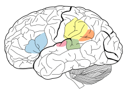
 Position of angular gyrus (shown in red).
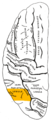 Lateral surface of left cerebral hemisphere, viewed from above. Angular gyrus is shown in orange.
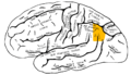 Lateral surface of left cerebral hemisphere, viewed from the side. Angular gyrus is shown in orange.
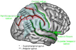
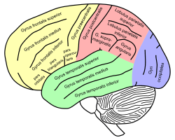 Lateral view of a human brain, main gyri labeled.
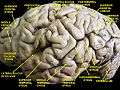 Cerebrum. Lateral view.Deep dissection.
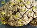 Cerebrum. Lateral view.Deep dissection.
References
- John, Hall (2010). Guyton and Hall Textbook of Medical Physiology. Saunders. p. 699. ISBN 978-1416045748.
- Park, HJ; Kim, JJ; Lee, SK; Seok, JH; Chun, J; Kim, DI; et al. (2008). "Corpus callosal connection mapping using cortical gray matter parcellation and DT-MRI". Human Brain Mapping. 29 (5): 503–16. doi:10.1002/hbm.20314. PMID 17133394.
- Makris, Nikos; Kennedy, David N.; McInerney, Sean; Sorensen, A. Gregory; Wang, Ruopeng; Verne, S. Caviness Jr; Pandya, Deepak N. (2005). "Segmentation of Subcomponents within the Superior Longitudinal Fascicle in Humans: A Quantitative, In Vivo, DT-MRI Study". Cereb. Cortex. 15 (6): 854–869. doi:10.1093/cercor/bhh186. PMID 15590909.
- Uddin, Lucina Q.; Supekar, Kaustubh; Amin, Hitha; Rykhlevskaia, Elena; Nguyen, Daniel A.; Greicius, Michael D.; Menon, Vinod (2010). "Dissociable Connectivity within Human Angular Gyrus and Intraparietal Sulcus: Evidence from Functional and Structural Connectivity". Cereb. Cortex. 20 (11): 2636–2646. doi:10.1093/cercor/bhq011. PMC 2951845. PMID 20154013.
- Rushworth, MF; Behrens, TE; Johansen-Berg, H (2006). "Connection patterns distinguish 3 regions of human parietal cortex". Cereb Cortex. 16 (10): 1418–1430. CiteSeerX 10.1.1.504.2825. doi:10.1093/cercor/bhj079. PMID 16306320.
- Makris, Nikos; Papadimitriou, George M.; Sorg, Scott; Kennedy, David N.; Caviness, Verne S.; Pandya, Deepak N. (2007). "The occipitofrontal fascicle in humans: A quantitative, in vivo, DT-MRI study". NeuroImage. 37 (4): 1100–1111. doi:10.1016/j.neuroimage.2007.05.042. PMC 3769215. PMID 17681797.
- Lee, H; Devlin, JT; Shakeshaft, C; Stewart, LH; Brennan, A; Glensman, J; Pitcher, K; Crinion, J; Mechelli, A; Frackowiak, RS; Green, DW; Price, CJ (2007). "Anatomical traces of vocabulary acquisition in the adolescent brain" (PDF). J Neurosci. 27 (5): 1184–1189. doi:10.1523/jneurosci.4442-06.2007. PMID 17267574.
- Ramachandran, V.S.; Hubbard, E.M (2003). "The Phenomenology of Synaesthesia" (PDF). Journal of Consciousness Studies. 10 (8): 49–57.
- Simon, K; Stilla, R; Sathian, K (2011). "Metaphorically feeling:Comprehending textual metaphors actives somatosensory cortex". Brain and Language. 120 (3): 416–421. doi:10.1016/j.bandl.2011.12.016. PMC 3318916. PMID 22305051.
- Brownsett, Sonia L. E.; Wise, Richard J. S. (2009-06-16). "The Contribution of the Parietal Lobes to Speaking and Writing". Cerebral Cortex. 20 (3): 517–523. doi:10.1093/cercor/bhp120. ISSN 1460-2199. PMC 2820696. PMID 19531538.
- Henschen, SL (1919). "On language, music and calculation mechanisms and their localisation in the cerebrum". Zeitschrift für die Gesamte Neurologie und Psychiatrie. 52: 273–298. doi:10.1007/bf02872428.
- Gerstmann, J (1940). "Syndrome of finger agnosia, disorientation for right and left, agraphia and acalculia—Local diagnostic value". Arch Neurol Psychiatry. 44 (2): 398–408. doi:10.1001/archneurpsyc.1940.02280080158009.
- Dehaene, S; Spelke, E; Pinel, P; Stanescu, R; Tsivkin, S (1999). "Sources of mathematical thinking: behavioral and brain-imaging evidence". Science. 284 (5416): 970–4. doi:10.1126/science.284.5416.970. PMID 10320379.
- Grabner, RH; Ansari, D; Reishofer, G; Stern, E; Ebner, F; Neuper, C (2007). "Individual differences in mathematical competence predict parietal brain activation during mental calculation". NeuroImage. 38 (2): 346–56. doi:10.1016/j.neuroimage.2007.07.041. PMID 17851092.
- Seghier, M. L. (2012). "The angular gyrus: multiple function ad multiple subdivisions". Neuroscientist. 19 (1): 43–61. doi:10.1177/1073858412440596. PMC 4107834. PMID 22547530.
- Arsalidou, M; Taylor, MJ (2011). "Is 2+2=4? Meta-analyses of brain areas needed for numbers and calculations". NeuroImage. 54 (3): 2382–93. doi:10.1016/j.neuroimage.2010.10.009. PMID 20946958.
- Hirnstein, M; Bayer, U; Ellison, A; Hausmann, M (2011). "TMS over the left angular gyrus impairs the ability to discriminate left from right". Neuropsychologia. 49 (1): 29–33. doi:10.1016/j.neuropsychologia.2010.10.028. PMID 21035475.
- Chen, Q; Weidner, R; Vossel, S; Weiss, PH; Fink, GR (Sep 2012). "Neural mechanisms of attentional reorienting in three-dimensional space". J Neurosci. 32 (39): 13352–62. doi:10.1523/jneurosci.1772-12.2012. PMID 23015426.
- Greicius, MD; Krasnow, B; Reiss, AL; Menon, V (2003). "Functional connectivity in the resting brain: a network analysis of the default mode hypothesis". Proc Natl Acad Sci U S A. 100 (1): 253–8. doi:10.1073/pnas.0135058100. PMC 140943. PMID 12506194.
- Farrer C, Frey SH, Van Horn JD, Tunik E, Turk D, Inati S, Grafton ST. The angular gyrus computes action awareness representations. Centre de Neuroscience Cognitive.
- Out-of-Body Experience? Your Brain Is to Blame - New York Times
- Arzy, S.; Seeck, M.; Ortigue, S.; Spinelli, L.; Blanke, O. (2006). "Induction of an illusory shadow person: Stimulation of a site on the brain's left hemisphere prompts the creepy feeling that somebody is close by". Nature. 443 (21): 287. doi:10.1038/443287a. PMID 16988702.
- Vallar G (July 2007). "Spatial neglect, Balint-Homes' and Gerstmann's syndrome, and other spatial disorders". CNS Spectr. 12 (7): 527–36. doi:10.1017/S1092852900021271. PMID 17603404.
- Carota A, Di Pietro M, Ptak R, Poglia D, Schnider A (2004). "Defective spatial imagery with pure Gerstmann's syndrome". Eur. Neurol. 52 (1): 1–6. doi:10.1159/000079251. PMID 15218337.
External links
| Wikimedia Commons has media related to Angular gyrus. |
- NIF Search - Angular Gyrus via the Neuroscience Information Framework
| Authority control |
|---|