Angioid streaks
Angioid streaks, also called Knapp streaks or Knapp striae are small breaks in Bruch's membrane, an elastic tissue containing membrane of the retina that may become calcified and crack.[1]
| Angioid streaks | |
|---|---|
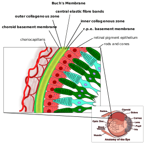 | |
| Bruchs membrane | |
| Specialty | Ophthalmology |
Clinical features
Angioid streaks are often associated with pseudoxanthoma elasticum, but have been found to occur in conjunction with other disorders, including Paget's disease, sickle cell disease and Ehlers-Danlos Syndrome. These streaks can have a negative impact on vision due to choroidal neovascularization or choroidal rupture. Also, vision can be impaired if the streaks progress to the fovea and damage the retinal pigment epithelium. The diagnosis is mainly clinical, however fundus fluorescene angiography shows that the streaks appear hyperfluorescent (window defect) in the early phase.
History
They were first described by Robert Walter Doyne in 1889 in a patient with retinal hemorrhages. A few years later, ophthalmologist Hermann Jakob Knapp called them "angioid streaks" because of their resemblance to blood vessels. From histopathological research in the 1930s, they were discovered to be caused by changes at the level of Bruch's membrane. Presently, it is believed that its pathology may be a combination of elastic degeneration of Bruch's membrane, iron deposition in elastic fibers from hemolysis with secondary mineralization, and impaired nutrition due to stasis and small vessel occlusion.